
- Title
-
A Temporal Chromatin Signature in Human Embryonic Stem Cells Identifies Regulators of Cardiac Development
- Authors
- Paige, S.L., Thomas, S., Stoick-Cooper, C.L., Wang, H., Maves, L., Sandstrom, R., Pabon, L., Reinecke, H., Pratt, G., Keller, G., Moon, R.T., Stamatoyannopoulos, J., and Murry, C.E.
- Source
- Full text @ Cell
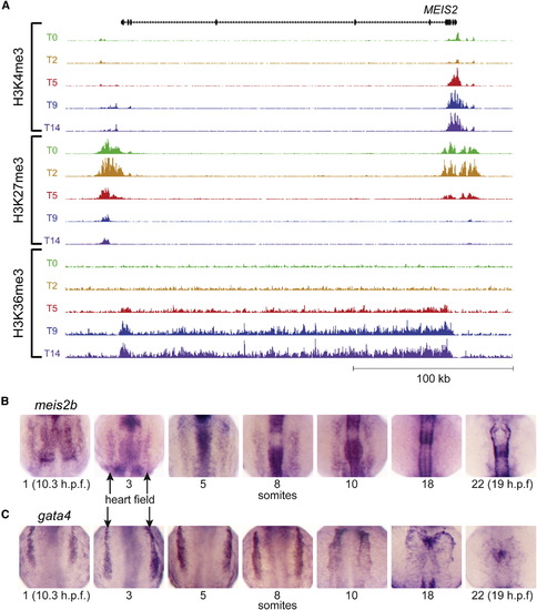
MEIS2 Chromatin Modifications during hESC Differentiation and Expression in Developing Zebrafish Embryos Resembles Other Regulators of Cardiac Development (A) The temporal pattern of epigenetic marks at the MEIS2 locus is similar to that of other regulators of cardiac development shown in Figure 1 (scales used: 1 to 250/150/25 tags per 150 bp for H3K4me3/H3K27me3/H3K36me3). (B) Zebrafish meis2b expression are shown for developing embryos at the 1, 3, 5, 8, 10, 18, and 22 somite stages, showing (C) similar expression patterns within the bilateral heart fields (arrows) to gata4 through the 10 somite stage. By 18 somites, meis2b is no longer expressed in the cardiac mesoderm whereas gata4 maintains expression. EXPRESSION / LABELING:
|
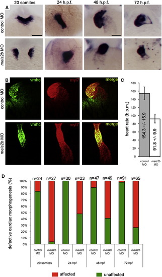
meis2b Is Required for Cardiac Morphogenesis (A) Expression of myl7 at 19 hpf, 24 hpf, 48 hpf, and 72 hpf in control-MO (top row) versus meis2b-MO (bottom row) injected zebrafish embryos. Dorsal view, anterior is up in 20 somite and 24 hpf embryos. Ventral view, anterior up in 48 hpf and 72 hpf embryos. At 19 hpf, meis2b-MO injected embryos display defects in fusion of the myl7+ cardiac progenitors at the midline compared with control-MO injected embryos. By 24 hpf, the heart tube has formed in meis2b morphants but displays aberrant cardiac morphogenesis and is either sitting at the midline or moving down the right side of the embryo, compared with the control-MO injected embryos where normal heart development proceeds with the heart tube emerging from under the head, down the left side of the embryo. At 48 and 72 hpf, control MO injected embryos display normal cardiac looping, whereas meis2b-MO injected embryos’ hearts have not looped. (B) This failure of cardiac looping in meis2b-MO injected embryos is further evident in vmhc (green) and myl7 (red) double fluorescent in situs at 48 hpf. (C) Heart rate is significantly reduced in meis2b-MO injected embryos compared with control-MO injected embryos at 72 hpf (b.p.m. = beats per minute) Mean heart rate ± SD is shown, n = 10. p = 4 × 10⁹ (Student’s t test, two-tailed). (D) Percentages of embryos displaying the depicted phenotypes. Scale bars, (A, top left), 100 μm, and (A, top third from the left), 50 μm. See also Figure S5. EXPRESSION / LABELING:
PHENOTYPE:
|
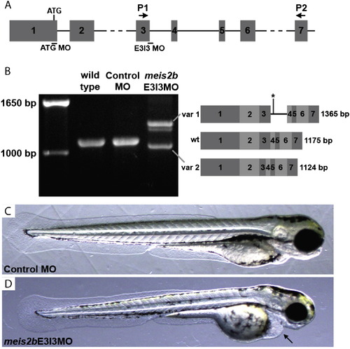
Validation of an Antisense Morpholino for Zebrafish meis2b Knockdown Shown in Figure 5 (A) Diagram of the first seven exons of zebrafish meis2b (NM_130910.1), shown approximately to scale, except for introns 2 and 6. Shown are the locations of binding sites for two antisense meis2b morpholinos, one targeting the start codon (ATG MO) to block translation, and one targeting the splice donor site for intron 3 (E3I3 MO) to block mRNA processing. Also shown are the locations of the primers (P1, P2) used in (B). (B) Reverse transcriptase polymerase chain reaction (RT-PCR) analysis of meis2b mRNA structure in wild-type, Standard control MO (control MO), and meis2bE3I3MO embryos. Sequencing of these RT-PCR products revealed wild-type and control MO embryos express wild-type meis2b transcript, while meis2bE3I3MO embryos express two transcript variants (diagrammed on the right). Variant 1 (var 1) retains intron 3, which encodes a premature stop codon (*) that occurs upstream of the Meis2b homeodomain. Variant 2 (var 2) results from aberrant splicing to an upstream cryptic splice donor, resulting in the loss of 51 base pairs of exon 3 but maintenance of the correct open reading frame in exon 4. (C and D) Live 72 hpf control MO and meis2bE3I3MO embryos. meis2bE3I3MO embryos show mild pericardial edema (arrow), as well as variable smaller heads and body curvature phenotypes. Anterior is to the right. PHENOTYPE:
|

Unillustrated author statements PHENOTYPE:
|
Reprinted from Cell, 151(1), Paige, S.L., Thomas, S., Stoick-Cooper, C.L., Wang, H., Maves, L., Sandstrom, R., Pabon, L., Reinecke, H., Pratt, G., Keller, G., Moon, R.T., Stamatoyannopoulos, J., and Murry, C.E., A Temporal Chromatin Signature in Human Embryonic Stem Cells Identifies Regulators of Cardiac Development, 221-232, Copyright (2012) with permission from Elsevier. Full text @ Cell