
|
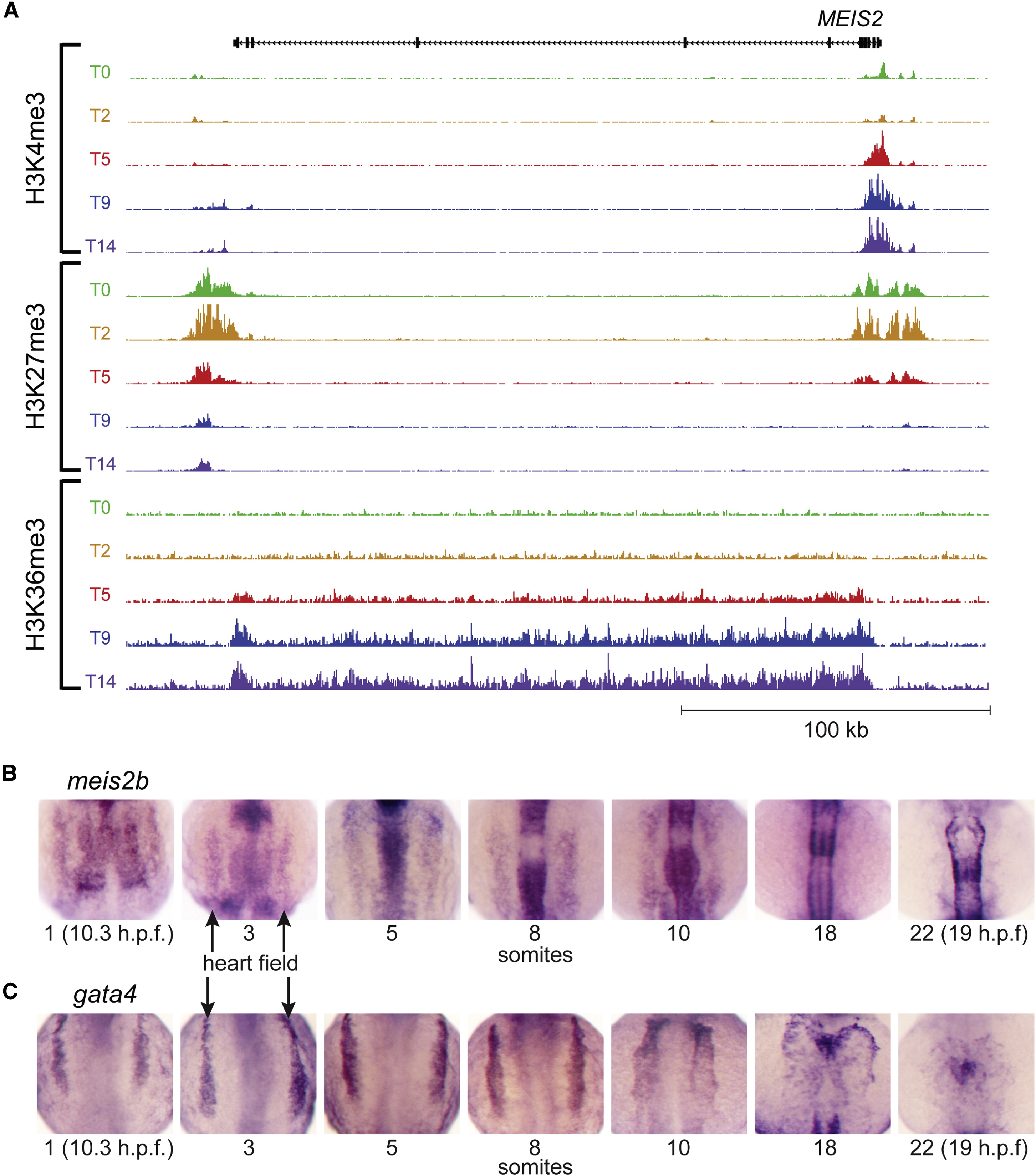
Fig. 4
MEIS2 Chromatin Modifications during hESC Differentiation and Expression in Developing Zebrafish Embryos Resembles Other Regulators of Cardiac Development (A) The temporal pattern of epigenetic marks at the MEIS2 locus is similar to that of other regulators of cardiac development shown in Figure 1 (scales used: 1 to 250/150/25 tags per 150 bp for H3K4me3/H3K27me3/H3K36me3). (B) Zebrafish meis2b expression are shown for developing embryos at the 1, 3, 5, 8, 10, 18, and 22 somite stages, showing (C) similar expression patterns within the bilateral heart fields (arrows) to gata4 through the 10 somite stage. By 18 somites, meis2b is no longer expressed in the cardiac mesoderm whereas gata4 maintains expression.
Reprinted from Cell, 151(1), Paige, S.L., Thomas, S., Stoick-Cooper, C.L., Wang, H., Maves, L., Sandstrom, R., Pabon, L., Reinecke, H., Pratt, G., Keller, G., Moon, R.T., Stamatoyannopoulos, J., and Murry, C.E., A Temporal Chromatin Signature in Human Embryonic Stem Cells Identifies Regulators of Cardiac Development, 221-232, Copyright (2012) with permission from Elsevier. Full text @ Cell