
- Title
-
Activity-based labeling of matrix metalloproteinases in living vertebrate embryos
- Authors
- Keow, J.Y., Pond, E.D., Cisar, J.S., Cravatt, B.F., and Crawford, B.D.
- Source
- Full text @ PLoS One
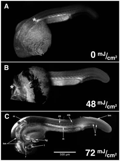
HxBP can be UV-photocrosslinked in vivo, and detected in situ by confocal microscopy. Composite confocal micrographs of 24 hpf zebrafish embryos injected anteriorly with 50 μM trifunctional HxBP probe, recovered, and exposed to increasing levels of UV irradiation reveals increasingly spatially structured patterns of fluorescence up to 72 mJ/cm2. Structures exhibiting strong HxBP labeling in 24 hpf embryos include the retina (r) and lens (l), head mesenchyme (hm), hatching gland (hg), perichordal sheath (ps), isolated notochord cells in the elongating region of the notochord (n), maturing myotome boundaries (mb), mesenchymal tissues of the trunk and tail (m), and the basement membrane underlying the epithelium dorsal to the elongating tail (bm). Asterisks mark the point of injection. |
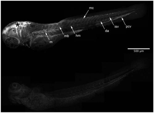
HxBP is labeling metalloproteinases in vivo. Composite confocal micrographs of 72 hpf zebrafish embryos injected anteriorly (*) with 50 μM of trifunctional HxBP probe either with (B) or without (A) competition from unlabeled GM6001. Embryos pre-injected with GM6001 show dramatically attenuated HxBP labeling, requiring the data shown in panel B to be collected using 42% increased gain in order to be detectable. Structures showing strong labeling in HxBP labeled 72 hpf embryos include proliferative myofibrils along the anterior lateral midline (m), maturing myotome boundaries (mb), the horizontal myoseptum (hm), individual migratory mesenchyme cells (mc), and the developing vasculature including the dorsal aorta (da), posterior cardinal vein (pcv) and intersomitic vessels (isv). Asterisk marks the injection site. Scale bar is 500 μM. |
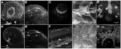
Details of HxBP labeling patterns reveal dynamics of active matrix remodeling. Z-projections of high magnification confocal micrographs illustrating patterns of HxBP labeling in both 24 hpf (A, B) and 72 hpf (C–H) zebrafish embryos. A) The developing zebrafish eye at 24 hpf, with HxBP labeling migratory mesenchyme (arrowhead), retinal epithelium (r), the choroid fissure and the hatching gland (hg). The lens shows no HxBP labeling, suggesting that matrix remodeling is absent in the lens. B) HxBP labeling the epithelial (e) and mesenchymal tissue (m) around the otic vesicle (ov), but no labeling within the otic vesicle. Primes are single focal planes of confocal stacks used to generate the non-prime panels. C) A ventrolateral view of the head of a 72 hpf embryo showing strong labeling in the developing scleral ossicles. Strong labeling is also evident in individual migratory mesenchyme cells and developing vasculature. D) lateral view of the anterior trunk showing strong labeling in the maturing myotome boundaries (mb), horizontal myoseptum (hm) and in individual myofibrils (mf) situated in the proliferative zone. E) Epithelia of the lateral head and dorsal aspect of the eye showing strong labeling in individual cells and patches of contiguous epithelial cells. F) lateral view of the tail showing strong labeling in migratory mesenchyme cells (mc), as well as labeling in the dorsal aorta (da), posterior cardinal vein (pcv) and intersomitic vessels (isv). G) lateral view of the surface epithelia covering the anterior trunk illustrating the ‘chicken wire’ patterning of HxBP labeling surrounding the periphery of the cells in this tissue. H) A dorsolateral view of the eye, illustrating strong HxBP labeling of mesenchymal cells invading across the surface of the retina and surrounding the lens (l). Scale bars are 50 μm in all panels. |

HxBP labeling in a 96 hpf swimming larva. Composite of confocal projections taken of a 96 hpf larva injected (at asterisk) with 50 μM trifunctional HxBP. Strong labeling is evident throughout the developing circulatory system, most notably in the looping vessels of the gill arches (g), the dorsal aorta (da), posterior cardinal vein (pcv), intersomitic vessels (isv), and hyaloid artery (ha). Labeling is also notable in individual migratory mesenchyme cells (mc), the protease-rich stomach (s), horizontal myoseptum (hm) and maturing craniofacial cartilages. Scale bar is 500 μm. |