
- Title
-
pitx2 Deficiency Results in Abnormal Ocular and Craniofacial Development in Zebrafish
- Authors
- Liu, Y., and Semina, E.V.
- Source
- Full text @ PLoS One
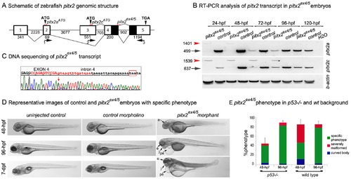
zebrafish pitx2 knockdown and associated phenotype. A. Schematic drawing of pitx2 genomic structure. Exons are shown as numbered boxes, sizes are indicated at the top (for exons) or at the bottom (for introns). The positions of primers to amplify pitx2 transcripts are shown and numbered 1–3; primers 1 and 3 are used for pitx2a and 2 and 3 for pitx2c. The position of antisense morpholino oligonucleotides, pitx2aATG, pitx2cATG and pitx2ex4/5sp, are shown with red lines. B. RT-PCR of pitx2 expression in pitx2ex4/5 morphants. Please note a complete absence of normal pitx2 transcripts (indicated with black arrows) and the presence of an abnormal large PCR product (indicated with red arrowheads) in mRNA extracted from pitx2ex4/5 embryos at 24–48-hpf, the presence of both normal (diminished) and abnormal products at 72–96-hpf pitx2ex4/5, and normal levels of pitx2 by 120-hpf due to weakening of morpholino effects. C. DNA sequencing of the abnormal PCR product observed in pitx2ex4/5 morphant embryos identified the presence of the 902-bp intron 4 in the pitx2ex4/5 transcript consistent with aberrant splicing (forward sequence is shown and the beginning of the intron is indicated with a red arrow; exon 4 sequence is shown in upper case while intron 4 is in lower case letters; the exon-intron junction sequence corresponding to the pitx2ex4/5 antisense oligomer is indicated in red). Therefore, the pitx2ex4/5 protein is predicted to contain partial pitx2 sequence (lacking amino acids encoded by exon 5) followed by 10 erroneous amino acids (pitx2ex4/5 stop codon is indicated with red box). D. Representative images of pitx2ex4/5 morphants, control morpholino-injected embryos and larvae developed from uninjected eggs at 48-hpf, 96-hpf and 7-dpf. E. Bar graph showing the distribution of observed embryonic phenotypes following pitx2ex4/5 morpholino injections into p53/ and wild-type zebrafish eggs. e- eye, j- jaw, pe- pericardial edema. |
zebrafish pitx2 knockdown and associated phenotype. A. Schematic drawing of pitx2 genomic structure. Exons are shown as numbered boxes, sizes are indicated at the top (for exons) or at the bottom (for introns). The positions of primers to amplify pitx2 transcripts are shown and numbered 1–3; primers 1 and 3 are used for pitx2a and 2 and 3 for pitx2c. The position of antisense morpholino oligonucleotides, pitx2aATG, pitx2cATG and pitx2ex4/5sp, are shown with red lines. B. RT-PCR of pitx2 expression in pitx2ex4/5 morphants. Please note a complete absence of normal pitx2 transcripts (indicated with black arrows) and the presence of an abnormal large PCR product (indicated with red arrowheads) in mRNA extracted from pitx2ex4/5 embryos at 24–48-hpf, the presence of both normal (diminished) and abnormal products at 72–96-hpf pitx2ex4/5, and normal levels of pitx2 by 120-hpf due to weakening of morpholino effects. C. DNA sequencing of the abnormal PCR product observed in pitx2ex4/5 morphant embryos identified the presence of the 902-bp intron 4 in the pitx2ex4/5 transcript consistent with aberrant splicing (forward sequence is shown and the beginning of the intron is indicated with a red arrow; exon 4 sequence is shown in upper case while intron 4 is in lower case letters; the exon-intron junction sequence corresponding to the pitx2ex4/5 antisense oligomer is indicated in red). Therefore, the pitx2ex4/5 protein is predicted to contain partial pitx2 sequence (lacking amino acids encoded by exon 5) followed by 10 erroneous amino acids (pitx2ex4/5 stop codon is indicated with red box). D. Representative images of pitx2ex4/5 morphants, control morpholino-injected embryos and larvae developed from uninjected eggs at 48-hpf, 96-hpf and 7-dpf. E. Bar graph showing the distribution of observed embryonic phenotypes following pitx2ex4/5 morpholino injections into p53/ and wild-type zebrafish eggs. e- eye, j- jaw, pe- pericardial edema. PHENOTYPE:
|
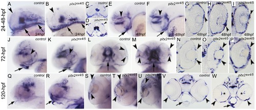
In situ hybridization with pitx2-exon 5 antisense riboprobe in control and morphant embryos. The pitx2 antisense riboprobe comprising exon 5 sequence detects wild-type and abnormally spliced pitx2ex4/5 transcripts. Images of whole mount embryos (A–F, J–M and Q–U) and sections (G–I, N–P and V, W) are shown for embryonic stages of 24-hpf (A–D), 48-hpf (E–I), 72-hpf (J–P) and 120-hpf (Q–W). Please note abnormal pitx2 transcripts around the developing eye (arrowheads in B, D) and pharyngeal arches (arrows in B) in morphants at 24-hpf, which is similar to pitx2 expression in control embryos (A, C). Staining for pitx2 positive cells in morphant embryos at 48-, 72- and 120-hpf identifies abnormal patterns during ocular and craniofacial development in comparison to pitx2 expression in control embryos (E–W). In terms of ocular patterns, some 48- and 72-hpf morphants demonstrate an accumulation of pitx2 transcriptionally active cells in the anterior segment of the eye (arrowheads; images for whole mount and sections from two different pitx2ex4/5 morphant embryos at 48-hpf (F, H, I) and 72-hpf (K, M, O, P) in comparison to control 48-hpf (E,G) and 72-hpf (J, L, N) are shown). In 120-hpf eyes, a disorganized pattern of pitx2 positive cells continues to be observed in pitx2ex4/5 morphant embryos (arrowheads in T, U, two different morphant embryos are shown, and W) in comparison to pitx2 expression in control embryos (S, V); in addition to the abnormal pattern in the anterior structures, an increased signal behind the lens corresponding to the hyaloid vasculature is also observed (asterisks in W). With regards to craniofacial development, in 72-hpf morphant embryos, strong staining is observed around the malformed oral cavity and arches with the level of expression similar to control pitx2 expression (arrows in J–M); at 120-hpf, pitx2 transcripts continue to be strongly expressed in the malformed pharyngeal arches (arrows in R and W) while expression of wild-type pitx2 is downregulated in control 120-hpf embryos (arrows in Q and V). |
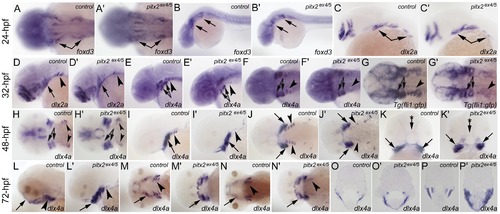
Craniofacial development and gene expression in pitx2ex4/5 morphants. Developmental patterns of foxd3, dlx2a and dlx4a, and Tg(fli1:gfp) expression in pitx2 ex4/5 embryos. In situ hybridization was performed with foxd3, dlx2a, dlx4a or gfp-specific antisense riboprobe in control (A–P) or morphant (A2–P2) zebrafish embryos. Please note similar expression patterns in migrating neural crest cells (foxd3) and pharyngeal arch primordial regions (dlx2a) at 24-hpf (arrows in control (A–C) and morphant (A2–C2) embryos). Starting at 32-hpf, expression of dlx2a, dlx4a and Tg(fli1:gfp) in the posterior arches appears to be reduced in pitx2ex4/5 fish (arrowheads in control (D–G) and morphant (D2–G2) embryos) while expression in the anterior arches (arrows in control (D–G) and morphant (D2–G2) embryos) is not significantly affected. In 48–72-hpf embryos, an increased expression of dlx4a in the anterior arches is detected (arrows in control (H–N) and morphant (H2–N2) embryos) while expression in the posterior arches remains reduced (arrowheads in control (H–N) and morphant (H2–N2) embryos); dlx4a expression around the oral cavity in 48-hpf embryos (arrow with asterisk in K and K2) as well as a frontward extension of the anterior arches at 72-hpf (arrows in L–N for controls and L2–N2 for morphants) are not observed in pitx2ex4/5 fish. Analysis of sections at 72-hpf also shows broadened expression of dlx4a in the craniofacial region of morphant embryos (O2, P2) in comparison to controls (O, P). EXPRESSION / LABELING:
|
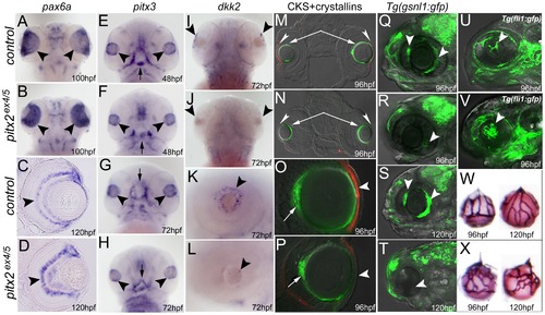
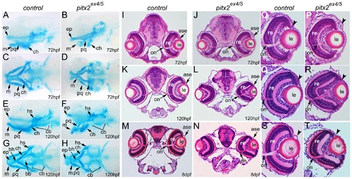
Ocular development and marker expression in pitx2ex4/5 morphants. In situ hybridization (A–L), double immunohistochemistry (M–P), live gfp fluorescence images (Q–V) and alkaline phosphatase staining (W, X) for control and morphant embryos are shown and are labeled with corresponding developmental stages in the lower right corner; dorsal (A, B, I, J) and ventral (E–H) views of a zebrafish larval head as well as transverse sections at the eye level (C, D, M–P), all positioned anterior to the top, and lateral views of an embryonic eye (K, L) or head (Q–V), positioned anterior to the left, are shown. Please note expression of pax6a in the developing retina of the control and morphant embryos (arrowheads in A–D); similar pitx3 expression in the developing lens in 48–72-hpf control and morphant embryos (arrowheads in E–H; abnormally developing pharyngeal arches and oral cavity in 48–72-hpf embryos are marked with arrows); reduced expression of dkk2 in the anterior segment of the pitx2 morphants (arrowheads in I–L); decreased/absent expression of corneal keratan sulfate proteoglycan (CKS) in the developing morphant corneas (red fluorescence, arrowheads in M–P) but similar expression of crystallins αA (green fluorescence, arrows in M, N) and βB1 (green fluorescence, arrows in O, P) in control and morphant lenses; and a decreased/absent expression of the Tg(gsnl1:gfp) transgene in the developing iridocorneal structures of pitx2ex4/5 morphants (arrowheads in R, T) in comparison to control embryos (Q, S). In addition to this, fluorescence (Tg(fli1:gfp) transgene) images (U, V) as well as alkaline phosphatase staining (W, X) demonstrate abnormal development of the hyaloid vasculature in pitx2ex4/5 morphants (arrowhead in V) in comparison to control embryos (U); increased number and disorganized appearance of pitx2ex4/5 hyaloid vessels (X) in comparison to controls (W) is revealed by alkaline phosphatase staining. |