
- Title
-
Evaluation of Sycp3, Plzf and Cyclin B3 expression and suitability as spermatogonia and spermatocyte markers in zebrafish
- Authors
- Ozaki, Y., Saito, K., Shinya, M., Kawasaki, T., and Sakai, N.
- Source
- Full text @ Gene Expr. Patterns
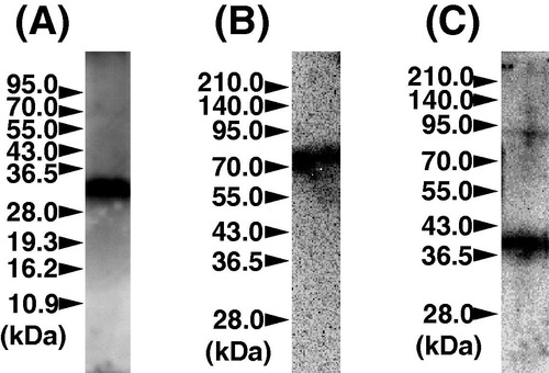
Western blot analysis of the zebrafish testis using antibodies against zebrafish Sycp3 (A), Plzf (B) and Cyclin B3 (C). EXPRESSION / LABELING:
|
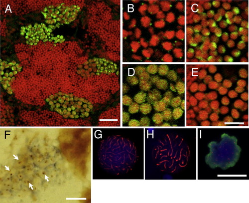
Expression of Sycp3 in the zebrafish testis by immunohistochemistry. (A-F) Sections of the zebrafish testis immunostained with anti-Sycp3 antibodies. Spermatocytes shown are at the preleptotene (B), leptotene (C), zygotene-pachytene (D) and diplotene (E) stages. Green signals indicate Sycp3 and red staining indicates nuclei. (F) A section of testis was hybridized with an anti-sense probe for sycp3 and then stained with anti-Sycp3 antibody. Dark blue color indicates sycp3 mRNA and brown color (arrowheads) indicates Sycp3 protein. (G-I) Chromosome spreads immunostained with anti-Sycp3 antibodies. Spermatocytes shown are at the zygotene (G), pachytene (H), and metaphase (I) stages. Red signals indicate Sycp3, blue staining indicates nuclei, and green staining indicates phosphohistone H3 (pH3, an M phase marker). Bars, 20 µm (A) and 10 µm (B-I). EXPRESSION / LABELING:
|
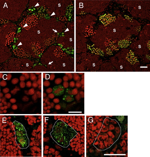
Expression of Plzf in the zebrafish testis by immunohistochemistry. (A) The section of a testis immunostained with antibodies against Plzf. Allows and arrowheads indicate type A spermatogonia and type B spermatogonia, respectively. Green signals indicate Plzf, and red staining indicates nuclei. S, sperm. (B) Immunostained with Sycp3 in the sequential section to (A). Green signals indicate Sycp3 and red staining indicates nuclei. S, sperm. (C and D) PI staining (C) and Plzf particles (D) of type A spermatogonia. (E-G) Immunostaining for Plzf in the 4-cell clone (E), the 8-cell clone (F), and the 16-cell clone (G). More than 10 clones of each were observed in two fish. White dots indicate surrounding Sertoli cells of the cyst. Bars, 20 µm (A, B, and E-G) and 10 µm (C and D). EXPRESSION / LABELING:
|
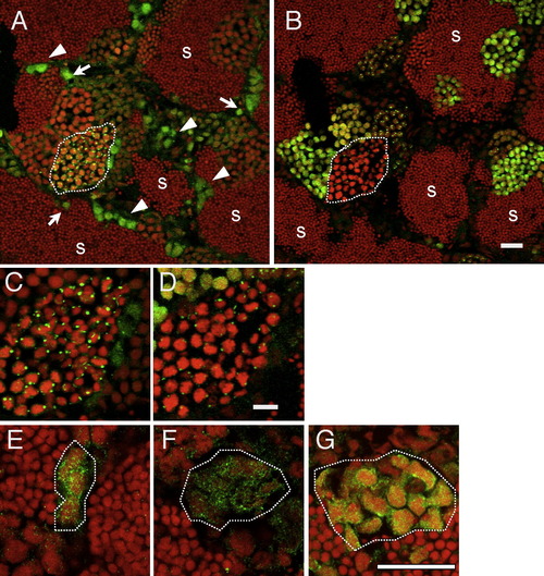
Expression of Cyclin B3 in the zebrafish testis by immunohistochemistry. (A and C) The section of a testis immunostained with antibodies against Cyclin B3. Allows and arrowheads indicate type A spermatogonia and type B spermatogonia, respectively. Green signals indicate Cyclin B3 and red staining indicates nuclei. S, sperm. (B and D) Immunostained with Sycp3 in the sequential section to (A) and (C). Green signals indicate Sycp3 and red staining indicates nuclei. S, sperm. Panels (C) and (D) are at higher magnification of the white dotted area in (A) and (B), respectively. (E-G) Immunostaining for Cyclin B3 in the 4-cell clone (E), the 8-cell clone (F), and the 16-cell clone (G). More than 10 clones of each were observed in two fish. White dots indicate surrounding Sertoli cells of the cyst. Bars, 20 µm (A, B, and E-G), 10 µm (C and D). EXPRESSION / LABELING:
|
Reprinted from Gene expression patterns : GEP, 11(5-6), Ozaki, Y., Saito, K., Shinya, M., Kawasaki, T., and Sakai, N., Evaluation of Sycp3, Plzf and Cyclin B3 expression and suitability as spermatogonia and spermatocyte markers in zebrafish, 309-315, Copyright (2011) with permission from Elsevier. Full text @ Gene Expr. Patterns