
|
Fig. 3
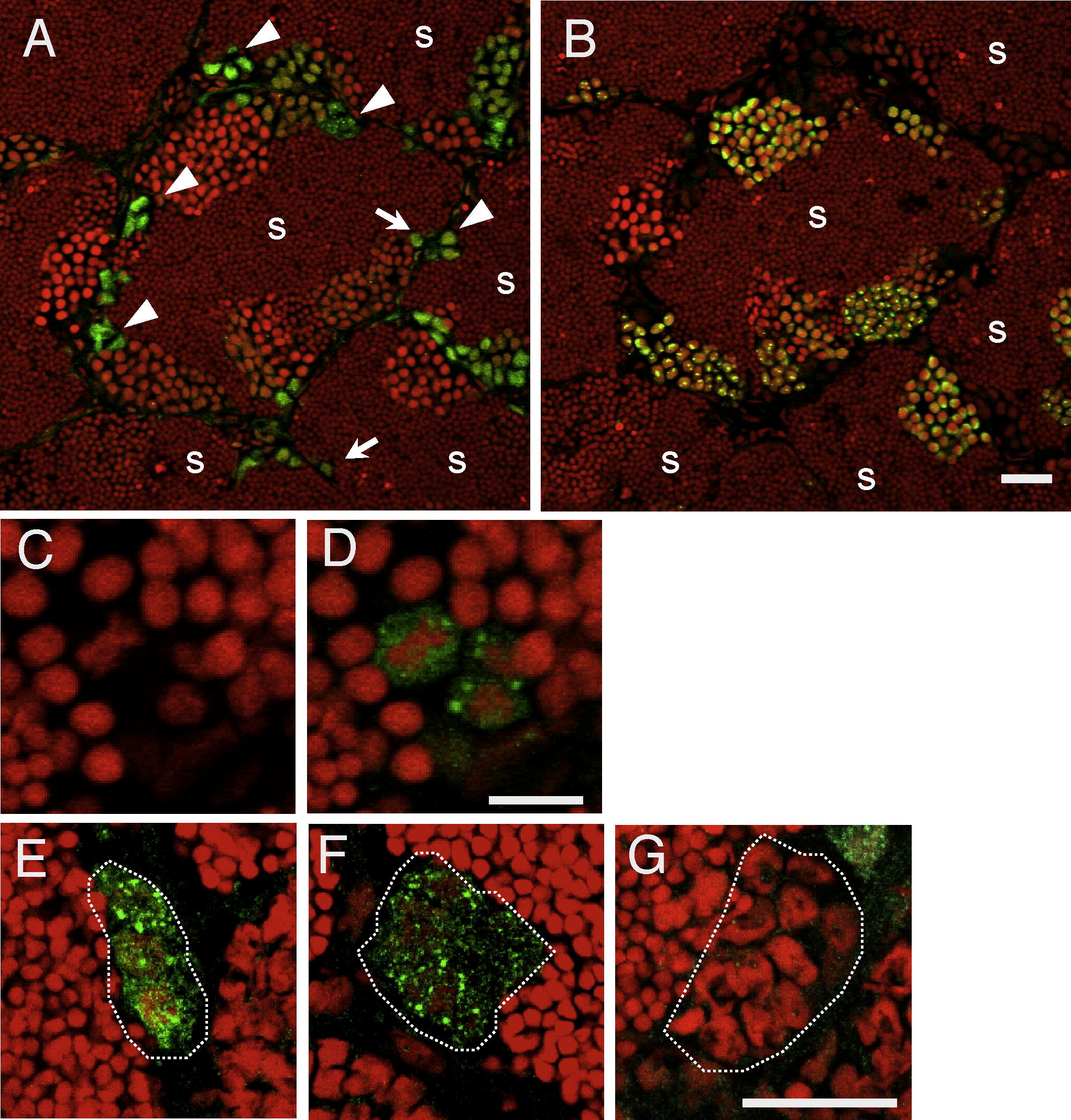
Expression of Plzf in the zebrafish testis by immunohistochemistry. (A) The section of a testis immunostained with antibodies against Plzf. Allows and arrowheads indicate type A spermatogonia and type B spermatogonia, respectively. Green signals indicate Plzf, and red staining indicates nuclei. S, sperm. (B) Immunostained with Sycp3 in the sequential section to (A). Green signals indicate Sycp3 and red staining indicates nuclei. S, sperm. (C and D) PI staining (C) and Plzf particles (D) of type A spermatogonia. (E-G) Immunostaining for Plzf in the 4-cell clone (E), the 8-cell clone (F), and the 16-cell clone (G). More than 10 clones of each were observed in two fish. White dots indicate surrounding Sertoli cells of the cyst. Bars, 20 µm (A, B, and E-G) and 10 µm (C and D).
Reprinted from Gene expression patterns : GEP, 11(5-6), Ozaki, Y., Saito, K., Shinya, M., Kawasaki, T., and Sakai, N., Evaluation of Sycp3, Plzf and Cyclin B3 expression and suitability as spermatogonia and spermatocyte markers in zebrafish, 309-315, Copyright (2011) with permission from Elsevier. Full text @ Gene Expr. Patterns