
|
Fig. 2
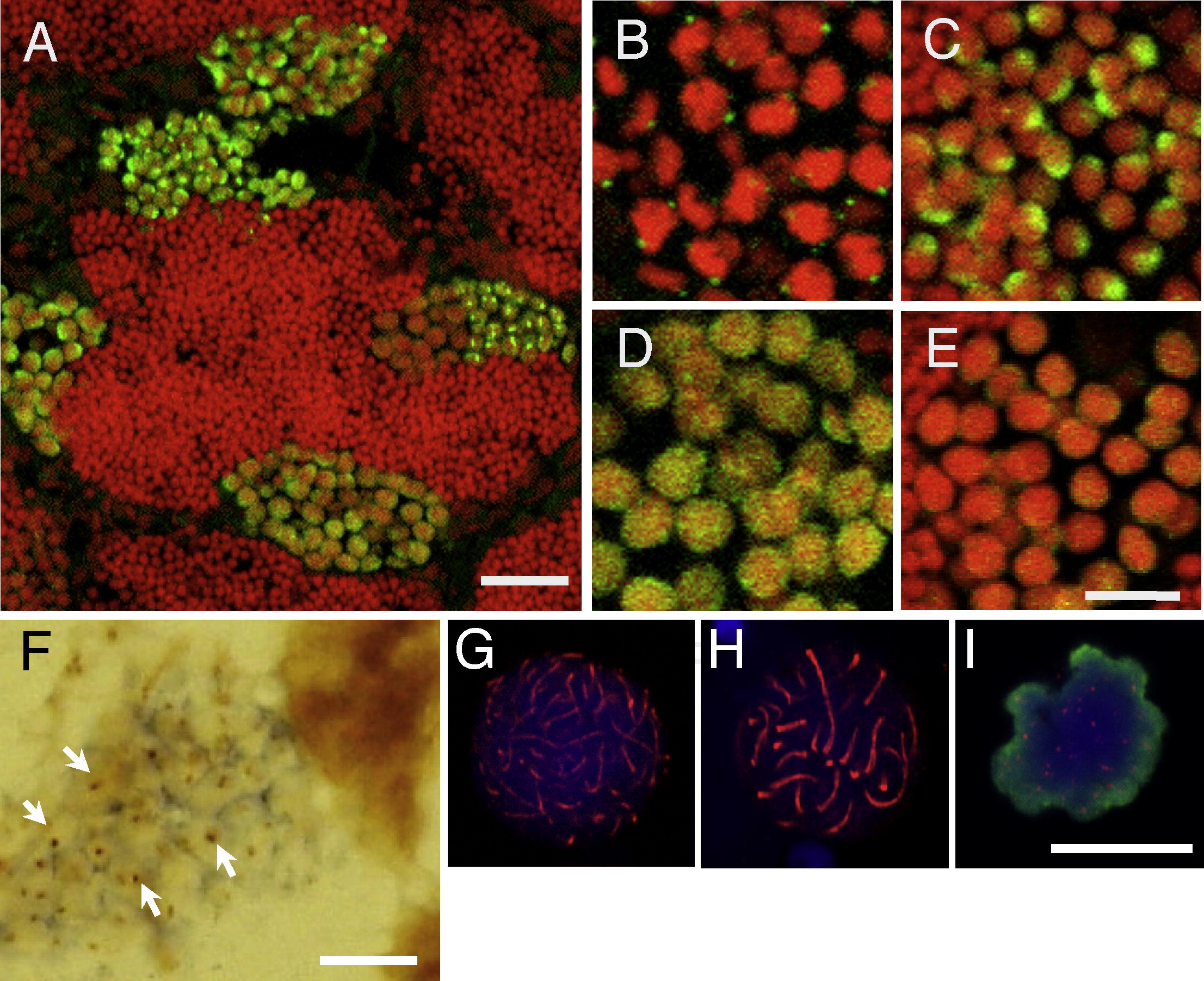
Expression of Sycp3 in the zebrafish testis by immunohistochemistry. (A-F) Sections of the zebrafish testis immunostained with anti-Sycp3 antibodies. Spermatocytes shown are at the preleptotene (B), leptotene (C), zygotene-pachytene (D) and diplotene (E) stages. Green signals indicate Sycp3 and red staining indicates nuclei. (F) A section of testis was hybridized with an anti-sense probe for sycp3 and then stained with anti-Sycp3 antibody. Dark blue color indicates sycp3 mRNA and brown color (arrowheads) indicates Sycp3 protein. (G-I) Chromosome spreads immunostained with anti-Sycp3 antibodies. Spermatocytes shown are at the zygotene (G), pachytene (H), and metaphase (I) stages. Red signals indicate Sycp3, blue staining indicates nuclei, and green staining indicates phosphohistone H3 (pH3, an M phase marker). Bars, 20 µm (A) and 10 µm (B-I).
Reprinted from Gene expression patterns : GEP, 11(5-6), Ozaki, Y., Saito, K., Shinya, M., Kawasaki, T., and Sakai, N., Evaluation of Sycp3, Plzf and Cyclin B3 expression and suitability as spermatogonia and spermatocyte markers in zebrafish, 309-315, Copyright (2011) with permission from Elsevier. Full text @ Gene Expr. Patterns