
- Title
-
prox1b Activity Is Essential in Zebrafish Lymphangiogenesis
- Authors
- Del Giacco, L., Pistocchi, A., and Ghilardi, A.
- Source
- Full text @ PLoS One
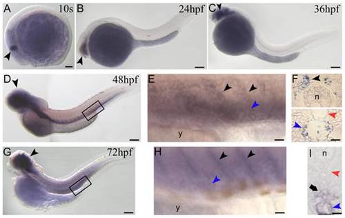
prox1b embryonic expression pattern analyzed by in-situ hybridization. (A) the first prox1b signal appears at 10 somite (s) stage in the central nervous system (CNS) (black arrowhead). (B) at 24hpf and (C) 36 hpf prox1b expression continues to be restricted to the CNS of the embryos (black arrowheads). (D) at 48 hpf prox1b mRNA is still detectable in the CNS, and is now expressed in the posterior cardinal vein (PCV) and the sprouts emanating from this vessel into the intersegmental space (black box). (E) magnification of the region boxed in D; PCV (blue arrowhead) and vein sprouts (black arrowheads) are indicated. (F) cross sections of 48 hpf-prox1b-hybridized embryos. (F, bottom) prox1b is expressed in the endothelial cells of the vein wall (blue arrowhead), and not in the dorsal aorta (DA) (red arrowhead). (F, top) prox1b signal is also visible on both sides of the trunk (black arrowhead), adjacent to the notochord (n). (G) at 72 hpf prox1b expression pattern is comparable to the 48 hpf stage, being the signal detectable in the CNS (black arrowhead), in the PCV and the sprouts emanating from the PCV (black box). (H) magnification of the region boxed in G; PCV (blue arrowhead) and vein sprouts (black arrowheads) are indicated. (I) cross section of a 72 hpf-prox1b-hybridized embryo. The endothelial cells of the PCV are still labeled (blue arrowhead), while the signal is completely absent from the DA territory (red arrowhead). prox1b is now expressed in the space comprised between the DA and the PCV (arrow), where the TD forms starting from around 72 hpf. y, yolk. Scale bars represent 100 μm (A,B,C,D,G) or 20 μm (E,F,H,I). EXPRESSION / LABELING:
|
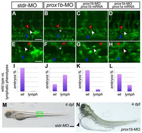
prox1b depletion results in the complete loss of the thoracic duct. fli1:GFP expression labels the DA (red arrowheads), the PCV (blue arrowheads), and lymphatic TD (white arrowheads) in 5 dpf embryos. A–D are shown magnified below in E-H. In comparison to control embryos injected with the stdr-MO (A,E), where 100% of the embryos analyzed displays a normal TD (I), the MO directed against prox1b (B,F) results in the absence of the TD (asterisks) in 70% of injected embryos (J). (C,G) the specificity of the effect of the prox1b-MO is confirmed by the ability of prox1b mRNA to rescue the lymphatic phenotype. (K) indeed, MO/mRNA coinjection resulted in about 90% of the embryos showing a normal TD. (D,H) prox1a mRNA is able to rescue the lymphatic phenotype induced by prox1b-MO injection in about 75% of the embryos analyzed (L). (M) 4 dpf control (stdr-MO) injected embryos compared to the (N) prox1b morphant (prox1b-MO) at the same developmental stage displaying severe edema (arrowhead). The green box in M indicates the approximate position of the embryo trunk depicted in A–D. Scale bars represent 40 μm (A,B,C,D), 20 μm (E,F,G,H), or 100 μm (M,N). |
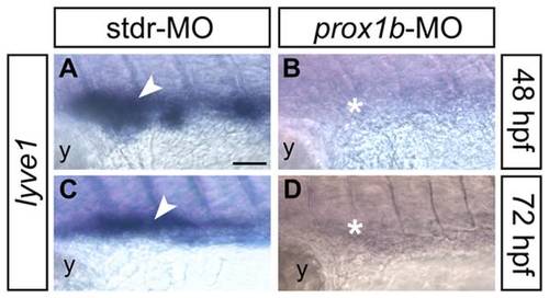
The expression of the lymphatic molecular marker lyve1 is dramatically reduced in prox1b morphants. lyve1 riboprobe labels developing lymphatic endothelial cells in 48 (A,B), and 72 (C,D) hpf embryos (all lateral views, anterior to the left; the approximate region of the embryo trunk imaged in all panels corresponds to the green box in Figure 3M). In comparison to control embryos injected with the stdr-MO (A,C), that display a normal expression of lyve1 (white arrowheads), the MO directed against prox1b (B,D) results in the absence of lyve1 signal (white asterisks). y, yolk. Scale bar represents 40 μm. EXPRESSION / LABELING:
PHENOTYPE:
|
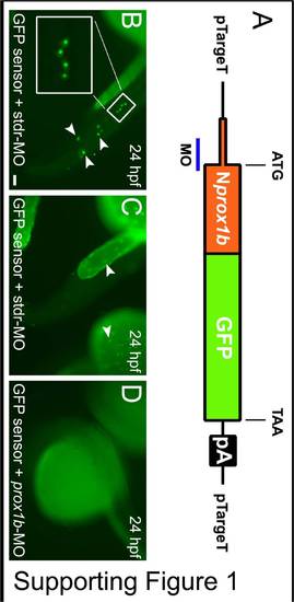
prox1b-MO specifically reduces prox1b mRNA translation. For the in-vivo test of the specificity of prox1b-MO, a prox1b-GFP sensor has been generated. (A) The construct contains 96 bp of the 52 UTR, and the first 432 bp of the prox1b coding sequence (Nprox1b) fused with the GFP open reading frame. The blue bar indicates the region of the mRNA targeted by the prox1b-MO. The construct, obtained by PCR, has been cloned into the pTargeT expression vector (Promega) and used for injection experiments. (B) GFP-positive cells in the trunk (inset and arrowheads) and (C) in the yolk epithelium (arrowheads) are visible following coinjection of the sensor and the stdr-MO. (D) The complete absence of fusion protein expression when the sensor is coinjected with prox1b-MO confirms the specificity of action of the morpholino. Scale bar represents 100 μm. |