
|
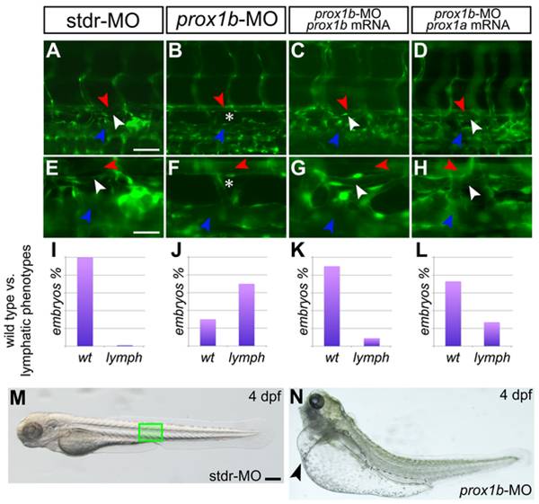
Fig. 3 prox1b depletion results in the complete loss of the thoracic duct.
fli1:GFP expression labels the DA (red arrowheads), the PCV (blue arrowheads), and lymphatic TD (white arrowheads) in 5 dpf embryos. A–D are shown magnified below in E-H. In comparison to control embryos injected with the stdr-MO (A,E), where 100% of the embryos analyzed displays a normal TD (I), the MO directed against prox1b (B,F) results in the absence of the TD (asterisks) in 70% of injected embryos (J). (C,G) the specificity of the effect of the prox1b-MO is confirmed by the ability of prox1b mRNA to rescue the lymphatic phenotype. (K) indeed, MO/mRNA coinjection resulted in about 90% of the embryos showing a normal TD. (D,H) prox1a mRNA is able to rescue the lymphatic phenotype induced by prox1b-MO injection in about 75% of the embryos analyzed (L). (M) 4 dpf control (stdr-MO) injected embryos compared to the (N) prox1b morphant (prox1b-MO) at the same developmental stage displaying severe edema (arrowhead). The green box in M indicates the approximate position of the embryo trunk depicted in A–D. Scale bars represent 40 μm (A,B,C,D), 20 μm (E,F,G,H), or 100 μm (M,N).