
- Title
-
Zebrafish calcium/calmodulin-dependent protein kinase II (cam-kii) inhibitors: Expression patterns and their roles in zebrafish brain development
- Authors
- Hsu, L.S., and Tseng, C.Y.
- Source
- Full text @ Dev. Dyn.
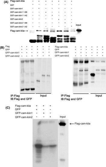
Zebrafish cam-kii inhibitors bound to and inhibited cam-kii α activity. A: The GST-pull down assay. The HEK 293 cells were transfected with pFlag-cam-kiiα plasmid. At 24 hr post-transfection, cell lysates were harvested and subjected to GST-pull down assay with GST or various C-terminal deleted forms of cam-kii inhibitors fusion protein beads as indicated in the presence of Ca2+/CaM. The pull-down complexes were subjected to Western blot analysis using anti-Flag antibody. Bottom: The equal amount of GST fusion proteins as visualized by Western blot analysis using anti-GST antibody. B: The co-immunoprecipitation assay. The HEK293 cells were co-transfected pFlag vector (left) or pFlag-cam-kiiα (right) combined with pEGFP, pEGFP-cam-kiin1, or pEGFP-cam-kiin2. At 24 hr post-transfection, cell lysates were harvested and subjected to immunoprecipitation with anti-Flag antibody. The immuno-complexes were probed with anti-GFP and anti-Flag antibody. C: The in vitro kinase assay. The HEK 293 cells were transfected with pFlag-cam-kiiα plasmid. At 24 hr post-transfection, cell lysates were immunoprecipitated with anti-Flag antibody. The immuno-complexes were subjected into kinase assay in the presence of Ca2+/CaM and GST, GST-cam-kiin1, or GST-cam-kiin2 fusion protein using myelin basic protein as substrate. These proteins were separated by electrophoresis and the phosphorylation was detected by autoradiography. Flag-cam-kiia denotes Flag-cam-kiiα. The data represent one of at least three independent experiments. |
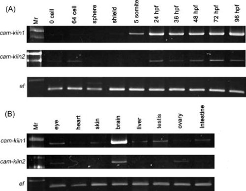
The spatial and temporal expression of cam-kii inhibitors by semi-quantitative analysis. The total RNA extracted from the indicated (A) stages and (B) adult tissues were subjected to RT-PCR analysis using the designed primers for cam-kiin1 and cam-kiin2. The elongation factor 1α (EF) was used as internal control. The PCR products were separated and visualized by ethidium bromide staining. Mr, 100-bp marker. |
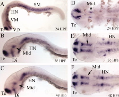
Expression patterns of (A-F) cam-kiin1 by whole mount in situ hybridization. Stages of embryos are indicated in the lower right. At 24 HPF, the cam-kiin1 was predominantly expressed in the telencephalon (T) region, ventral diencephalons (VD), ventral midbrain (VM), and somites (SM). At 36 and 48 HPF, cam-kiin1 was localized in telencephalon (T), diencephalons (Di), midbrain (Mid), and neurons in hindbrain (HN). A-C: Lateral view; D-F: flattened dorsal view. All images are anterior to the left. |
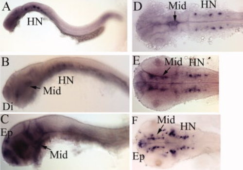
Expression patterns of (A-F) cam-kiin2 by whole mount in situ hybridization. The cam-kiin2 RNA was shown to be wildly expressed in brain region with stronger in neurons in hindbrain (HN) at 24. At 36 HPF, cam-kiin2 appeared in diencephalon (Di), midbrain (Mid), and neurons in hindbrain (HN). At 48 HPF, RNA transcripts of cam-kiin2 were detected in epiphysis (Ep), diencephalon (Di), midbrain (Mid), and neurons in hindbrain (HN). A-C: Lateral view; D-F: flattened dorsal view. All images are anterior to the left. |
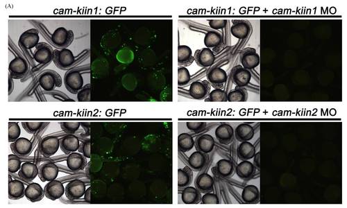
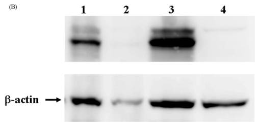
Specific blocked of GFP reporter expression by cam-kii inhibitors MO. A: Two GFP reporter plasmids (cam-kiin1: gfp and cam-kiin2: gfp) that encompassed the MO target sequences were constructed. Embryos that received 100 pg cam-kiin1: gfp or cam-kiin2: gfp resulted in mosaic GFP expression at 24 HPF. Co-injection of cam-kiin1: gfp or cam-kiin2: gfp with corresponding MO significantly diminished GFP expression. Left panel in each group: the gross morphology was pictured under light contrast microscopy. Right panel in each group: the expression of GFP was detected under fluorescent microscopy. B: Cell lysates derived from embryos injected with cam-kiin1: gfp (lane 1), cam-kiin1: gfp + cam-kiin1 MO (lane 2), cam-kiin2: gfp (lane 3), and cam-kiin2: gfp + cam-kiin2 MO (lane 4) were subjected to Western blot analysis using anti-GFP antibody. Significantly decreased GFP expression was detected in lane 2 and 4. β-actin served as internal control. |
|
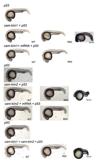
Morphological phenotype of cam-kii inhibitors morphants. Embryos that received 10 ng p53, cam-kiin1, or cam-kiin2, or combined both plus p53 MO were examined at 24 hr postfertilization (HPF). Embryos injected with p53 MO exhibited normal development. cam-kiin1 or cam-kiin2 or combined both plus p53 morphants were subdivided into wild type and mild phenotype as described in the text. PHENOTYPE:
|
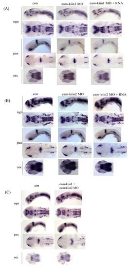
Characterization of (A) cam-kiin1, (B) cam-kiin2, and (C) double knockdown morphants. Expression of ngn1 (ngn), pax2.1 (pax), and otx2 (otx) in embryos that received morpholino with or without corresponding mRNA at 24 HPF were analyzed. Top: ngn1 and pax 2.1: lateral view; bottom: ngn1 and pax 2.1 and otx2: flattened dorsal view. All images are anterior to the left. |

Unillustrated author statements EXPRESSION / LABELING:
|