
- Title
-
Characterization of zebrafish intestinal smooth muscle development using a novel sm22{alpha}-b promoter
- Authors
- Seiler, C., Abrams, J., and Pack, M.
- Source
- Full text @ Dev. Dyn.
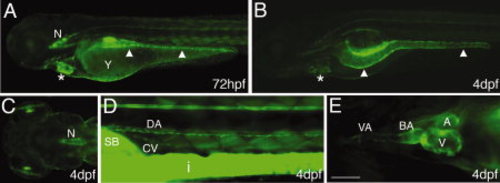
ECR5 of the sm22α-b promoter can drive GFP expression in smooth muscle cells. At 72 hpf (A), expression is detectable in the intestine (arrowheads) heart (*), the rostral notochord (“N”), and trunk muscle cells. Starting at 4 dpf (B-E), expression is present in the intestine (arrowheads B), in the notochord (“N”, C), the dorsal aorta (“DA” D), and the cardinal vein (“CV” D). In the heart (E), GFP is expressed in the bulbus arteriosus (“BA”), the atrium (“A”), and the ventricle (“V”) and the ventral aorta (“VA”). A, B, D, lateral view; C, dorsal; E, ventral. Scale bar in E = 250 7mu;m in A-C, 100 μm in D, and 50 μm in E. EXPRESSION / LABELING:
|
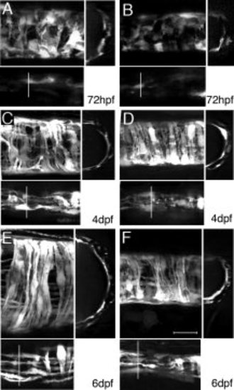
Smooth muscle differentiation. GFP expression in smooth muscle of the middle (A, C, E) and posterior (B, D, F) intestine. A-F: Large panel: maximum intensity projections of confocal scans through half of the intestine (lateral view). Lower panels: top 15 μm to visualize longitudinal layer; right panels: z-section at the position indicated in the lower panel. The longitudinal cell layer is located on top of the circular layer but is less bright and thus appears below in the large panels. At 72 hpf (A, B), circular layer cells are present but appear undifferentiated, with a round morphology and projections to adjacent cells. Few undifferentiated longitudinal layer cells are present (A, B, lower panel). At 4dpf (C, D) and 6dpf (E, F), cells become increasingly directional and differentiate to long strands. Both outer longitudinal and inner circular layers are distinguishable. Scale bar in F = 20 μm for all panels. EXPRESSION / LABELING:
|
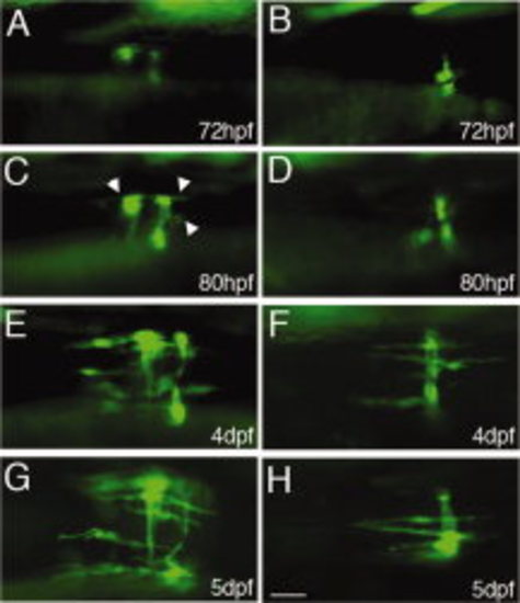
Clonal analysis of smooth muscle development. Mosaic expression of the ECR5 -GFP transgene (Tg(sm22αb: GFP)) in developing smooth muscle cells in a domain in the middle of the intestine (A, C, E, G) and the posterior intestine (B, D, F, H). Frequently patches of 2-3 cells can be found at 72 hpf (A, B). At 80 hpf, the cells elongate in circular directions and the first longitudinal layer cells become visible in the middle domain (arrowheads in C). More longitudinal layer cells develop between 80 and 96 hpf. They are always located close to circular layer cells. Scale bar in H = 25 μm for all panels. |
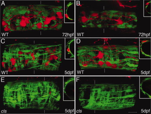
Development of enteric nervous system (ENS) and smooth muscle. 3D reconstruction of smooth muscle cells expressing GFP (green) and ENS cells expressing DsRed (red) (lateral view; insets: z-sections at position indicated by broken line). Middle (A,C,E) and posterior (B,D,F) intestine at 72 hpf (A, B) and 5 dpf (C-F). Enteric neurons with well-defined axonal projections are detected at 72 hpf while the smooth muscle is still differentiating (A, B) and the cells have a round shape in the posterior (B). At 72 hpf, ENS cells are positioned above the smooth muscle cells (A, B). At 5dpf, most ENS cells are located above the circular layer within the longitudinal smooth muscle layer (C, D). Axons can often been seen in parallel to longitudinal layer cells. Smooth muscle differentiation appears normal in colourless (cls) mutant larvae without ENS (E, F). Scale bar in F = 20 μm for all panels. EXPRESSION / LABELING:
|