
|
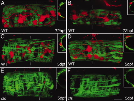
Fig. 5 Development of enteric nervous system (ENS) and smooth muscle. 3D reconstruction of smooth muscle cells expressing GFP (green) and ENS cells expressing DsRed (red) (lateral view; insets: z-sections at position indicated by broken line). Middle (A,C,E) and posterior (B,D,F) intestine at 72 hpf (A, B) and 5 dpf (C-F). Enteric neurons with well-defined axonal projections are detected at 72 hpf while the smooth muscle is still differentiating (A, B) and the cells have a round shape in the posterior (B). At 72 hpf, ENS cells are positioned above the smooth muscle cells (A, B). At 5dpf, most ENS cells are located above the circular layer within the longitudinal smooth muscle layer (C, D). Axons can often been seen in parallel to longitudinal layer cells. Smooth muscle differentiation appears normal in colourless (cls) mutant larvae without ENS (E, F). Scale bar in F = 20 μm for all panels.