|
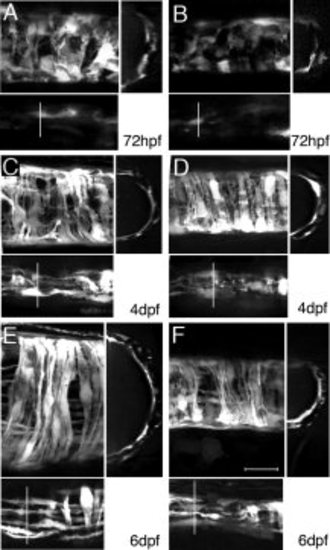
Smooth muscle differentiation. GFP expression in smooth muscle of the middle (A, C, E) and posterior (B, D, F) intestine. A-F: Large panel: maximum intensity projections of confocal scans through half of the intestine (lateral view). Lower panels: top 15 μm to visualize longitudinal layer; right panels: z-section at the position indicated in the lower panel. The longitudinal cell layer is located on top of the circular layer but is less bright and thus appears below in the large panels. At 72 hpf (A, B), circular layer cells are present but appear undifferentiated, with a round morphology and projections to adjacent cells. Few undifferentiated longitudinal layer cells are present (A, B, lower panel). At 4dpf (C, D) and 6dpf (E, F), cells become increasingly directional and differentiate to long strands. Both outer longitudinal and inner circular layers are distinguishable. Scale bar in F = 20 μm for all panels.
|