
- Title
-
Heartbeat regulates cardiogenesis by suppressing retinoic acid signaling via expression of miR-143
- Authors
- Miyasaka, K.Y., Kida, Y.S., Banjo, T., Ueki, Y., Nagayama, K., Matsumoto, T., Sato, M., and Ogura, T.
- Source
- Full text @ Mech. Dev.
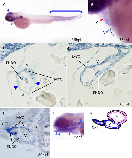
Expression patterns of miR-143. (A) miR-143 is expressed in the brain (blue arrowhead), heart (red arrowhead) and somites of the posterior body part (blue bracket) at 36 hpf. (B) In a magnified view, expression is relatively restricted in the ventricular domain (V, red arrowhead). Atrial expression is not evident at this stage (A, blue arrowhead). The outline of the atrium is demarcated by a dotted line. (C and D) Ventricular expression (indicated by the letter V) is detected in both the myocardium (MYO) and the endocardium (ENDO). Expression is not observed in the atrial compartment (indicated by the letter A). The position of the A–V junction is indicated by blue arrowheads in C. (E) At 80 hpf, robust expression is evident in the OFT, while the ventricular expression (indicated by V) is less intense, and the endocardial expression is weak. miR-143 is not expressed in atrial regions (indicated by A). (F) At 5 dpf, the OFT expression becomes intense (red arrowhead), while the surrounding expression is detected in the head chondrocytes (blue arrowheads) and the branchial arches (blue bracket). (G) A schematic representation of miR-143 expression. miR-143 is expressed in a gradient manner, with the highest signals in the OFT and less intense ones in the ventricular myocardium. Neither endocardial nor myocardial expression is detected in the atrial compartment. |
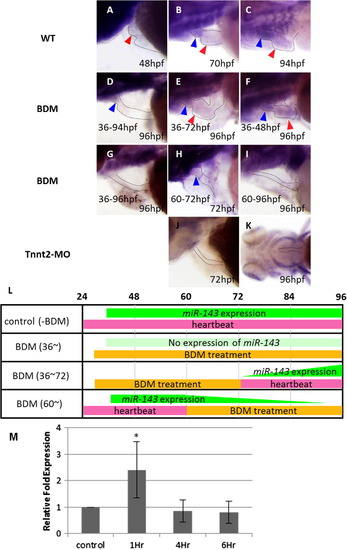
Heartbeat dependency of miR-143 expression. (A–C) Expression of miR-143 in the ventricle (red arrowheads) and the OFT (blue arrowheads) becomes evident as heartbeat and blood circulation increase. (D) Heartbeat was arrested from 36 hpf by BDM, then restarted by washing out at 94 hpf. Only faint expression of miR-143 was detected at 96 hpf in the OFT (blue arrowhead). (E) Heartbeat was stopped from 36 to 72 hpf. Continuous beating from 72 to 96 hpf allowed weak expression of miR-143 both in the OFT and the ventricle. (F) Duration of heartbeat for 48 h restored the expression of miR-143. (G) Heartbeat was arrested from 36 to 96 hpf. Expression of mir-143 was not detected at 96 hpf. (H) When heartbeat was stopped from 60 to 72 hpf, only faint expression of miR-143 was observed in the OFT at 72 hpf (blue arrowhead). (I) 36 h arrest of heartbeat (from 60 to 96 hpf) abolished expression completely. (J and K) In Tnnt2 morphants, expression of miR-143 was not detected at both 72 (J) and 96 hpf (K). (L) A schematic representation of miR-143 expression. Normally, heartbeat and blood circulation initiate at 24 hpf. Expression of miR-143 commences soon after at 36 hpf. When heartbeat was stopped from 36 hpf, miR-143 was not expressed. When BDM was washed out to initiate heartbeat at 72 hpf, miR-143 was induced. Likewise, when heartbeat was arrested at 60 hpf, miR-143 expression was decreased. (M) Expression of miR-143 was quantified by quantitative RT-PCR in human umbilical vein endothelial cells (HUVECs), to which 2 Pa (20 dyne/cm2) of shear stress was applied. miR-143 was induced rapidly within 1 h. This induction was transient and returned to a basal level within 4 h. |
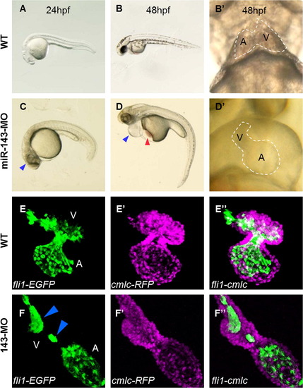
Cardiac phenotypes of the miR-143 morphants. At 24 hpf, miR-143 morphants showed a hypoplastic brain (blue arrowhead) (C), which were not observed in the WT (A and B). (D) At 48 hpf, inflation of the epicardial sac (blue arrowhead) with stagnation of red blood cells in the yolk sac (red arrowhead) was evident. (D′) At 48 hpf, the atrium (indicated by the letter A) was enlarged, and the ventricle (indicated by the letter V) failed to expand in the morphants, in contrast to the two chambered WT heart (B′). (E) In the normal heart, the endocardial lining was visualized by EGFP, which was driven by the endocardial cell-specific fli1 promoter. In the morphants, only the atrial part was enlarged, leaving the ventricular domain tubular (F). In such heart, the lining of endocardial cells was irregular and interrupted, showing a string-like arrangement (blue arrowheads). (E′ and F′) Cardiomyocytes were visualized by RFP, which was driven by the cardiomyocyte-specific cmlc promoter. |
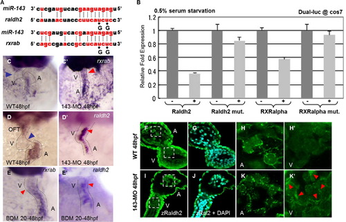
miR-143 regulates RA signaling. (A) An alignment of the sequences of miR-143 and its targets, raldh2 and rxrab. (B) Dual luciferase assay was performed with Cos7 cells cultured in the presence of 0.5% of serum. Expression of miR-143 repressed the reporters that contain the target motifs of raldh2 and rxrab (bars indicated by +), whereas the repressive effect of miR-143 was cancelled by mutating their sequences as shown in panel (A) (indicated by Raldh2 mut and RXRalpha mut). In the WT heart, rxrab (C) and raldh2 (D) were expressed in the atrial endocardium, not in the ventricular endocardium (blue arrowheads). In the morphants, both rxrab (C′) and raldh2 (D′) were ectopically induced in the ventricular endocardium (red arrowheads). When the beating of the WT hearts was stopped by BDM from 20 to 48 hpf, both rxrab (E) and raldh2 (E′) were induced in the ventricular compartment (red arrowheads), like the morphants. (F–K′) Raldh2 protein was detected by an antibody against it. In the WT, cytoplasmic staining was observed only in the atrial endocardial cells (F and G). Areas indicated by dotted lines were magnified in (H and H′). Raldh2 protein localizes in the cytoplasm of the atrial endocardium (H), not in the ventricular endocardium (H′). In the morphants, raldh2 was detected in both the atrial and ventricular endocardium (I and J). In magnified views, weak but distinct staining of raldh2 was observed even in the ventricular compartment (K′). EXPRESSION / LABELING:
|

Myocardial phenotypes of the miR-143 morphants. (A) Phalloidin staining revealed thick actin bundles formed along the atrio-ventricular axis (blue arrowheads). (B–B′′′) In a magnified view, such bundles were pan-Cadherin-negative (blue arrowheads in B′), indicating that they were formed beyond the boundaries of cardiomyocytes. White dotted lines indicate the position of the bundles. (C to D′′′) In the miR-143 morphants, the thick longitudinal actin bundles were not formed. Rather, bundles were formed along the boundaries of cardiomyocytes, as visualized by pan-Cadherin (D′′) and DAPI staining (D′′′). Note the dot-like aggregation of the pan-Cadherin signals (D′′). (E and G) In both the WT and the miR-143 morphants, atrial phalloidin staining showed a similar mesh-like structure. (F) In the WT embryos, the amhc gene is expressed exclusively in the atrial myocardium. (H) In the miR-143 morphants, amhc is ectopically expressed in the atrium (red arrowheads). (I) Tissue stiffness of the ventricles and the atrium was measured separately by a microtensile tester. The ventricle of the WT heart was stiffer than the WT atrium (gray bars). In contrast, stiffness of the morphant ventricles decreased to the level of the WT atrium, and the morphant atrium was also affected, yet in a lesser extent (light gray bars). |
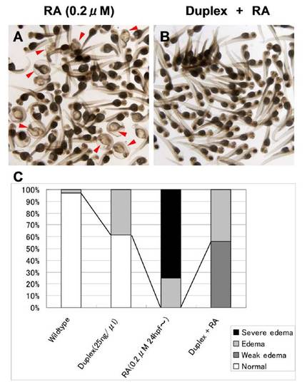
(A) As a combination of gain-of-function and rescue experiments, we soaked zebrafish embryos in a saline solution containing 0.2μM of RA from 24 to 60 hpf. Approcimately 75% of RA-treated embryos displayed severe edema in the epicardial sacs (red arrowheads). (B) When a duplex of miR-143 was injected, RA-induced edema was reduced to only mild to weak edema. (C) Occurrence of the edematous phenotype in the heart. Injection of the miR-143 duplez resulted in the edematous phenotype in all embryos, with severe epicardial edema in more than 70% of cases. When the RA treatment and the injection of the duplex were combined, this phenotype became milder. |
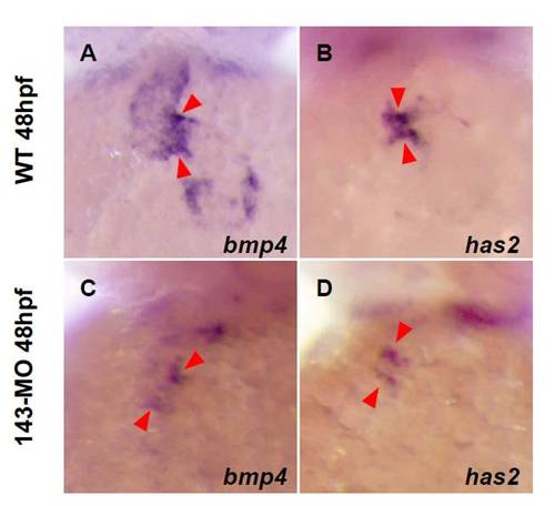
(A and B) Expression patterns of EXPRESSION / LABELING:
|
Reprinted from Mechanisms of Development, 128(1-2), Miyasaka, K.Y., Kida, Y.S., Banjo, T., Ueki, Y., Nagayama, K., Matsumoto, T., Sato, M., and Ogura, T., Heartbeat regulates cardiogenesis by suppressing retinoic acid signaling via expression of miR-143, 18-28, Copyright (2011) with permission from Elsevier. Full text @ Mech. Dev.