
|
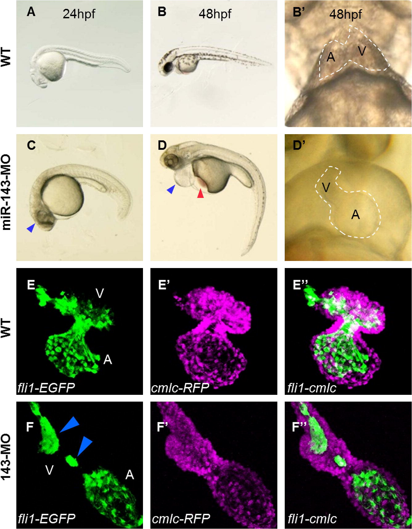
Fig. 3 Cardiac phenotypes of the miR-143 morphants. At 24 hpf, miR-143 morphants showed a hypoplastic brain (blue arrowhead) (C), which were not observed in the WT (A and B). (D) At 48 hpf, inflation of the epicardial sac (blue arrowhead) with stagnation of red blood cells in the yolk sac (red arrowhead) was evident. (D′) At 48 hpf, the atrium (indicated by the letter A) was enlarged, and the ventricle (indicated by the letter V) failed to expand in the morphants, in contrast to the two chambered WT heart (B′). (E) In the normal heart, the endocardial lining was visualized by EGFP, which was driven by the endocardial cell-specific fli1 promoter. In the morphants, only the atrial part was enlarged, leaving the ventricular domain tubular (F). In such heart, the lining of endocardial cells was irregular and interrupted, showing a string-like arrangement (blue arrowheads). (E′ and F′) Cardiomyocytes were visualized by RFP, which was driven by the cardiomyocyte-specific cmlc promoter.
Reprinted from Mechanisms of Development, 128(1-2), Miyasaka, K.Y., Kida, Y.S., Banjo, T., Ueki, Y., Nagayama, K., Matsumoto, T., Sato, M., and Ogura, T., Heartbeat regulates cardiogenesis by suppressing retinoic acid signaling via expression of miR-143, 18-28, Copyright (2011) with permission from Elsevier. Full text @ Mech. Dev.