
- Title
-
A direct role for Wnt8 in ventrolateral mesoderm patterning
- Authors
- Baker, K.D., Ramel, M.C., and Lekven, A.C.
- Source
- Full text @ Dev. Dyn.
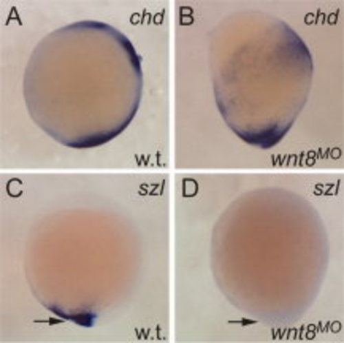
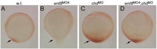
wnt8 morphants have reduced BMP signaling. A-D: In situ hybridizations on bud stage embryos, lateral views, anterior up. A: chordin expression marks several domains in the wild-type that are expanded in wnt8 loss of function embryos (B). C: sizzled expression is a readout for BMP signaling in the tail at bud stage (arrow). D: This expression is absent in wnt8 morphants. |
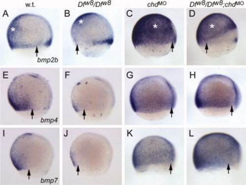
Wnt8 is not required for BMP expression. In situ hybridizations at -70% epiboly to detect expression of bmp2b (A-D), bmp4 (E-H), and bmp7 (I-L). Shown are lateral views, anterior up, dorsal right. Genotypes are indicated above each row. A,E,I: bmp2b, bmp4, and bmp7 are expressed in the ventral embryo. Arrows indicate the dorsal limit of the observable signal for each ligand. B,F,J: Ventral BMP expression is significantly reduced in wnt8 mutant embryos, indicated by ventral shift in position of arrow. Ventral ectoderm bmp2b expression is not as significantly affected (white asterisk). C,G,K: Chordin knockdown leads to dorsal expansion of BMP expression. Note dorsal shift of arrow positions. D,H,L: BMP ligand expression is dorsally expanded in wnt8 mutants that have Chordin knocked down. Note the expanded ventral ectoderm domain of bmp2b in wnt8;chordin double loss-of-function (asterisk in D), compared to wnt8 mutant (asterisk in B). While bmp2b in the margin does not appear to recover in the double loss-of-function (arrow in D), both bmp4 and bmp7 show dorsally expanded expression in the double loss-of-function. |
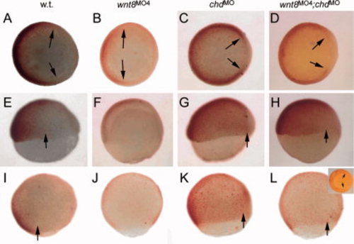
Wnt8 is not required for BMP signaling activity. Images of anti-phosphorylated Smad1/5 staining. Genotypes are indicated above each row. Arrows indicate dorsal extent of visible labeling. A-D: Animal pole views of shield stage embryos, dorsal right. E-H: Lateral views, shield stage, dorsal right. I-L: Lateral views, -80% epiboly, anterior up, dorsal right. P-Smad1/5 is confined to the ventral embryo at shield stage (A,E), and expression is significantly reduced in wnt8 morphants (B,F). P-Smad1/5 expands dorsally (arrows) in chordin morphants (C,G) and in wnt8;chordin morphants (D,H). P-Smad1/5 can still be detected in a dorsally expanded domain of chordin (K) and wnt8;chordin (L) morphants in late gastrulation. Inset in L: vegetal view, dorsal right; arrows indicate dorsal extent of staining. Note absence of staining in wnt8 morphant at 80% epiboly (J). |
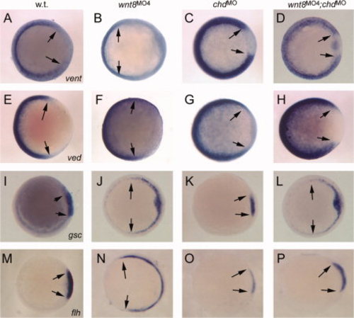
Differential organizer regulation by Wnt8 and BMP signaling. In situ hybridizations for vent (A-D), ved (E-H), gsc (I-L), and flh (M-P). All images show animal pole views, shield stage, dorsal right. Arrows indicate detectable limits of corresponding expression domain. Knockdown condition is indicated above each column. Note expansion of vent and ved in double knockdown (D,H), which correlates with rescued flh expression (P) but not gsc (L). |
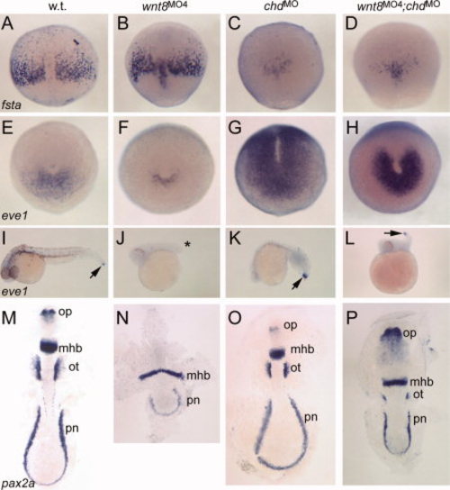
Differential response of ventrolateral mesoderm to Wnt8 and BMP regulation. In situ hybridizations to fsta (A–D), eve1 (E-L), and pax2a (M-P). A-D: 80% epiboly, dorsal view, anterior up. fsta is slightly expanded in wnt8 morphants, but is suppressed in chordin morphants (C) and wnt8;chordin double morphants (D). E-H: Tailbud stage, posterior view, dorsal up. eve1 marks tailbud progenitors. Note reduced expression in wnt8 morphant (F), but strongly expanded expression in chordin morphant (G) and somewhat expanded expression in wnt8;chordin morphants (H). I-L: -30 hpf, lateral views, anterior left. eve1 continues to mark tailbud progenitors (I, arrow). Note absence in wnt8 morphant (J, asterisk) but presence in chordin morphant (K) and wnt8;chordin double morphant (L, arrow). M-P: Five- to six-somite stage, flat mount, anterior up. pax2a labels optic stalk (op), midbrain-hindbrain boundary (mhb), otic vesicle (ot), and pronephros (pn). Note absence of optic stalk and otic vesicle staining in wnt8 morphant (N) and slight narrowing of neural plate in chordin morphant (O). In the double morphant (P), note posterior expansion of optic stalks, slightly widened MHB, reduced otic vesicles, and shortened pronephros domain. |
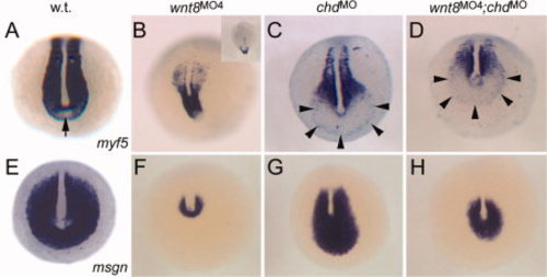
Wnt8 and BMP input on tailbud progenitor specification. All images: posterior views, dorsal up, 5-6 somite stage. A–D: myf5 expression marks presomitic mesoderm and somites, but is excluded from a small region of the ventral tailbud (arrow in A). Expression is significantly reduced in wnt8 morphant (B; inset: dorsal view). Note expansion of myf5-free progenitor zone in chordin morphant (C, outlined by arrowheads), and smaller expansion in wnt8;chordin morphants (D). E-H: msgn expression mirrors the behavior of the myf5-free zone of the tailbud. |
Wnt8 is not required for BMP signaling activity after gastrulation. Images of anti-phosphorylated Smad1/5 staining at the 5-6-somite stage. Genotypes are indicated above each row. Arrows indicate tail domain where nuclear P-Smad1/5 indicates active BMP signaling. Lateral views, dorsal right, anterior up. Note absence of staining in tailbud of wnt8 morphant (B), elevated staining in chordin morphant (C), and strong staining in wnt8;chordin double morphant (D). |
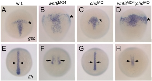
Differential dorsal mesoderm regulation by Wnt8 and BMP signaling. In situ hybridizations on bud stage embryos to detect gsc (A-D) and flh (E-H). Genotypes are indicated above each column. Asterisks in A-D indicate prechordal plate mesoderm domain. Note expansion in wnt8 and wnt8;chordin morphants (B,D), but not chordin morphant (C). Arrows in E-H indicate width of notochord domain. Note expansion in wnt8 morphant (F) but not chordin or wnt8;chordin morphants (G,H). These results show that the observations made at shield stage (see Fig. 4) continue to hold true through gastrulation. |
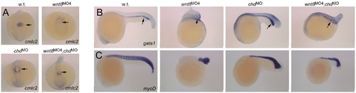
Differential response of additional ventrolateral mesoderm domains to Wnt8 and BMP regulation. In situ hybridizations at 24 hpf to detect cmlc2 in the heart (A), gata1 in blood progenitors (B), and myoD in body musculature (C). A: Dorsal views of the anterior embryo, anterior pointing down. Arrows indicate cmlc2-expressing cardiac cells. Note near-absence of staining in wnt8 morphant, but presence in chordin and wnt8;chordin morphants. B: Lateral views, anterior left. Arrows indicate gata1-expressing blood progenitors. Note severe reduction in wnt8 morphant, expansion in chordin morphant and mild reduction in wnt8;chordin morphant. C: Lateral views, anterior left. myoD expression marks somitic muscles. Note reduction of somites in wnt8 morphant, increased myoD expression in the most posterior region of chordin morphant, and additive phenotype in wnt8;chordin morphant. These results mirror our observations of pax2a expression domains as shown in Figure 5, i.e., defects of mesodermal patterning observed in wnt8 morphants is rescued by elevated BMP signaling, but posterior growth of tissues is not. |
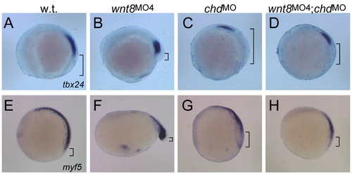
Alternate view of the tailbud and presomitic mesoderm in wnt8, chordin, and wnt8;chordin morphants. All images are lateral views, 5-6-somite stage embryos, anterior to left, dorsal up. Genotypes are indicated above each column. Brackets indicate the tailbud, defined as tissue posterior to presomitic mesoderm. A-D: tbx24 is expressed in presomitic mesoderm. Note the strong reduction in tailbud tissue observed in wnt8 morphants (B), significant expansion in chordin morphants (C), and approximately wild-type size observed in wnt8;chordin morphant (D). E-H: Lateral views of embryos stained for myf5 expression reveal a similar result. Note that myf5 expression is also in adaxial cells that flank the notochord into the tailbud, thus the apparent size of the tailbud in this view is smaller than that observed in tbx24-stained embryos. This is thus an optical effect, yet the result is not different between the two probes used. |