
|
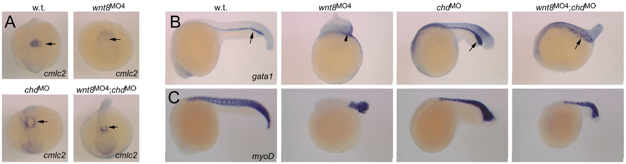
Fig. S3 Differential response of additional ventrolateral mesoderm domains to Wnt8 and BMP regulation. In situ hybridizations at 24 hpf to detect cmlc2 in the heart (A), gata1 in blood progenitors (B), and myoD in body musculature (C). A: Dorsal views of the anterior embryo, anterior pointing down. Arrows indicate cmlc2-expressing cardiac cells. Note near-absence of staining in wnt8 morphant, but presence in chordin and wnt8;chordin morphants. B: Lateral views, anterior left. Arrows indicate gata1-expressing blood progenitors. Note severe reduction in wnt8 morphant, expansion in chordin morphant and mild reduction in wnt8;chordin morphant. C: Lateral views, anterior left. myoD expression marks somitic muscles. Note reduction of somites in wnt8 morphant, increased myoD expression in the most posterior region of chordin morphant, and additive phenotype in wnt8;chordin morphant. These results mirror our observations of pax2a expression domains as shown in Figure 5, i.e., defects of mesodermal patterning observed in wnt8 morphants is rescued by elevated BMP signaling, but posterior growth of tissues is not.