
Fig. 5
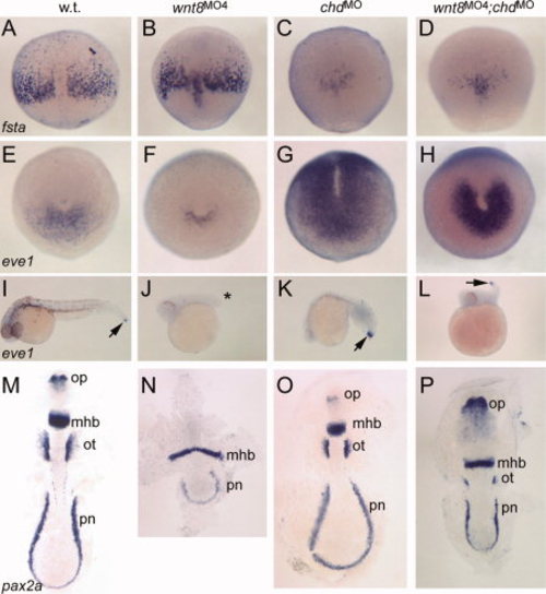
Differential response of ventrolateral mesoderm to Wnt8 and BMP regulation. In situ hybridizations to fsta (A–D), eve1 (E-L), and pax2a (M-P). A-D: 80% epiboly, dorsal view, anterior up. fsta is slightly expanded in wnt8 morphants, but is suppressed in chordin morphants (C) and wnt8;chordin double morphants (D). E-H: Tailbud stage, posterior view, dorsal up. eve1 marks tailbud progenitors. Note reduced expression in wnt8 morphant (F), but strongly expanded expression in chordin morphant (G) and somewhat expanded expression in wnt8;chordin morphants (H). I-L: -30 hpf, lateral views, anterior left. eve1 continues to mark tailbud progenitors (I, arrow). Note absence in wnt8 morphant (J, asterisk) but presence in chordin morphant (K) and wnt8;chordin double morphant (L, arrow). M-P: Five- to six-somite stage, flat mount, anterior up. pax2a labels optic stalk (op), midbrain-hindbrain boundary (mhb), otic vesicle (ot), and pronephros (pn). Note absence of optic stalk and otic vesicle staining in wnt8 morphant (N) and slight narrowing of neural plate in chordin morphant (O). In the double morphant (P), note posterior expansion of optic stalks, slightly widened MHB, reduced otic vesicles, and shortened pronephros domain. |