
- Title
-
The X-linked mental retardation gene SMCX/JARID1C defines a family of histone H3 lysine 4 demethylases
- Authors
- Iwase, S., Lan, F., Bayliss, P., de la Torre-Ubieta, L., Huarte, M., Qi, H.H., Whetstine, J.R., Bonni, A., Roberts, T.M., and Shi, Y.
- Source
- Full text @ Cell
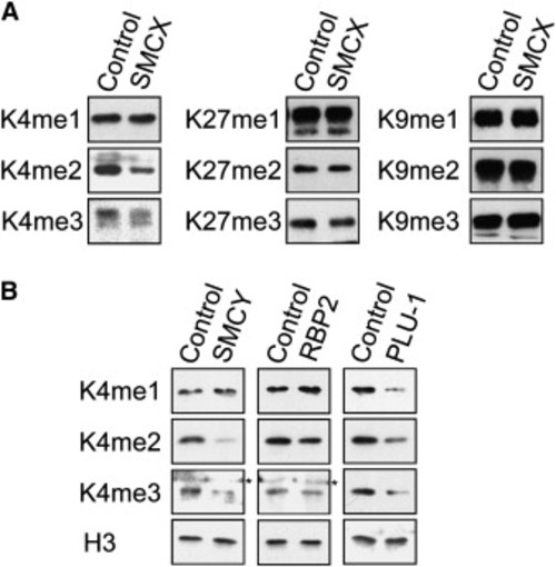
Specific Demethylation of Methylated H3K4 on Native Histone by SMCX, SMCY, Plu-1, and RBP2 (A and B) Calf thymus histones (3 μg) were incubated with 3 μg of purified, full-length SMCX, SMCY, RBP2, and PLU-1, respectively, and subjected to western blot analysis using antibodies that specifically recognize methylated histones. Heat-inactivated recombinant proteins were used as controls. Reduced signals were found only for H3K4me3 and H3K4me2 by SMCX and SMCY and for all three methylation states by Plu-1. Nonspecific signals were marked as asterisks. |
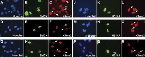
Overexpression of SMCX Resulted in Reduction of H3K4me2/3 Signals In Vivo Expression construct carrying HA-SMCX (A–I) or a mutant HA-SMCX (H514A, J–R) was transiently transfected into U2OS cells and stained by anti-HA (green) and appropriate antibodies against methylated histones (H3K4me1, H3K4me2, or H3K4me3; red). Nucleus was counterstained by Hoechst 33342. Comparing to untransfected cells, significant decrease of K4me2 and K4me3 level was found in SMCX overexpressing cells (C and F, arrows). The demethylation activity was completely abrogated by H514A mutation (O and R, arrows). Overexpression of neither wild-type SMCX nor SMCX (H514A) affected the K4me1 status (C and L, arrows). |
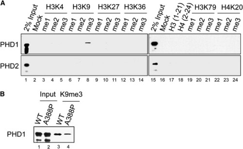
Recognition of H3K9me3 by a PHD Finger of SMCX (A) Histone-peptide binding assay. Two PHD fingers of SMCX, referred as PHD1 and PHD2, were fused to GST and purified from bacteria. Biotinylated histone peptides shown in a panel were incubated with purified PHD fingers, and pulled down by streptavidin agarose. After washing, the bound GST fusion proteins were visualized by western blot analysis using anti-GST antibody. Among the differently methylated histone peptides that we tested, only H3K9me3 peptide bound PHD1. No detectable binding was found for PHD2. (B) A388P mutation associated with X-linked mental retardation affects PHD1 binding to H3K9me3. GST-fused wild-type and mutant PHD1 carrying A388P were incubated with H3K9me3 peptide and washed four times, and the bound fractions were analyzed by western blot analysis. |

Brain-Specific Expression and a Role for Smcx in Neuronal Survival during Zebrafish Development (A) RNA in situ hybridization of whole-mount zebrafish embryos. In (a–f), antisense smcx RNA probe hybridized with embryos at the time points indicated. Arrows point to the neural tube, and arrowheads indicate muscles that were weakly stained. In (g–l), or the control, sense smcx probe hybridized with embryos of the same developmental stages. (B) Smcx is required for proper neuronal development and survival. Zebrafish embryos injected with two independent antisense morpholinos (MO 1 and 2) to smcx show developmental delay and have abnormal brain patterning (black arrowheads) at 24 hr postfertilization (hpf) compared with embryos injected with a control morpholino. At 48 hpf, knockdown embryos show moderate ventral curvature of the tail. Acridine orange (AO) staining at 30 hpf reveals dramatic cell death throughout the brain (white arrowheads) and neural tube (white arrows) in knockdown embryos. (C) Transplantation of MO-treated cells to wild-type embryos. Live cell bodies labeled with green dye in the head of most morpholino-treated cells are visible, and often long neuronal processes can be observed (b). Most smcx MO-treated cells with red dye formed small diffuse spots, likely to be dead cell aggregates (c). Bright field view is shown in (a). EXPRESSION / LABELING:
|
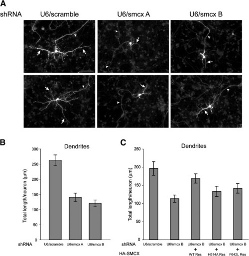
Smcx Is Required for Dendritic Development in Rat Cerebellar Granule Neuron (A) Representative images of primary cultured rat cerebellar granule neurons, which were cotransfected with the U6/scramble, U6/smcxA, or U6/smcxB shRNA plasmid and a GFP expression vector and subjected to immunocytochemistry with a GFP antibody. Arrows and arrowheads indicate dendrites and axons, respectively. Scale bar denotes 50 μm. (B) Analysis of dendritic length of neurons transfected in (A) shows that knockdown of Smcx led to a significant decrease in total dendritic length with two different RNA hairpins (p < 0.0001; ANOVA followed by Bonferonni-Dunn posthoc test, 160 neurons measured). Data presented as mean ± SEM. (C) SMCX-dependent dendritic morphogenesis is linked to SMCX demethylase activity. Primary granule neurons were transfected with U6/scramble or U6/smcx B RNAi plasmid, together with a control expression vector or an expression plasmid encoding the RNAi-resistant human SMCX protein (WT Res) or point mutants that disrupt demethylase activity (H514A Res and F642L Res). Dendritic length was significantly reduced in Smcx knockdown neurons as compared to U6/scramble-transfected neurons (p < 0.0001; ANOVA followed by Bonferonni-Dunn posthoc test). Dendritic length was significantly increased in neurons expressing WT Res SMCX, but not in H514A Res- or F642L Res-expressing neurons, in the background of Smcx RNAi, as compared to Smcx RNAi alone (p < 0.0041; ANOVA followed by Bonferonni-Dunn posthoc test). Total number of neurons measured was 265. Data presented as mean ± SEM. |
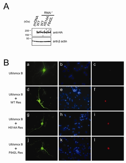
Comparable Expression of RNAi-Resistant SMCX in Rat Cerebellar Granule Neurons |
Reprinted from Cell, 128(6), Iwase, S., Lan, F., Bayliss, P., de la Torre-Ubieta, L., Huarte, M., Qi, H.H., Whetstine, J.R., Bonni, A., Roberts, T.M., and Shi, Y., The X-linked mental retardation gene SMCX/JARID1C defines a family of histone H3 lysine 4 demethylases, 1077-1088, Copyright (2007) with permission from Elsevier. Full text @ Cell