
- Title
-
Zebrafish as a Model for Cancer Self-Renewal
- Authors
- Ignatius, M.S., and Langenau, D.M.
- Source
- Full text @ Zebrafish
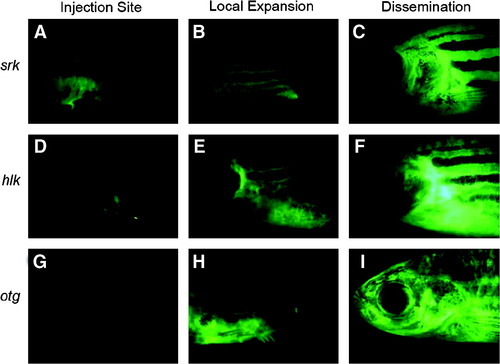
Heritable T-cell malignancies are transplantable and can be easily visualized by fluorescent protein expression in leukemic cells (modified from Frazer et al.41). Green fluorescent protein (GFP+) leukemias from shrek (srk; A–C), hulk (hlk; D–F), and oscar the grouch (otg; G–I) mutants were transplanted into irradiated wild-type hosts. At 1 week, GFP+ cells were seen at the site of transplantation (A, D, G). Subsequently, tumors spread locally (B, E, H) and then disseminated widely (C, F, I). |
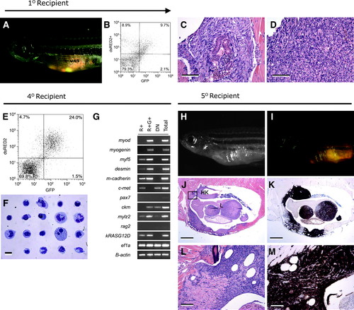
The dsRED2+ cell population from double transgenic rag2-dsRED2/alpha-actin-GFP animals contains the serially transplantable cancer stem cell in zebrafish embryonal rhabdomyosarcoma (ERMS). (A–D) Primary transplanted tumors from alpha-actin-GFP+/rag2-dsRED2+ fish (1° Recipient). (A) Merged image of GFP fluorescent, dsRED2 fluorescent, and bright field images. (B) Fluorescence-activated cell sorting (FACS) analysis of primary recipient engrafted with ERMS. (C, D) Histological analysis revealed heterogeneity in transplant animals, with some fish having masses of spindled cells (C) or round cell aggregates (D), or both. Scale bars equal 100μm in (C, D). (E–G) Cells isolated from serially transplanted animals, in this case a quaternary recipient animal (4° Recipient). (E) FACS plot of tumor cells isolated from a 4° recipient. (F) Wright-Giemsa-stained cytospins of FACS-sorted R+ cells from quaternary tumor. (G) Semiquantitative reverse transcription–polymerase chain reaction analysis of FACS-sorted cell populations. Total refers to total cells isolated from quaternary transplanted ERMS isolated by FACS based on cell viability and serve as an input control. (H–M) Fish transplanted with 50 R+ cells defined in (E)–(G) (5° Recipient). (H) Brightfield image of transplant recipient animal. (I) Merged image of a dsRED2+/GFP+ tumor in same animal. (J) Hematoxylin and eosin-stained and (K) anti-GFP-immunostained section of transplanted fish showing that ERMS cells infiltrate the liver (L), head kidney (HK), and skeletal muscle. (L, M) High-power magnification of boxed region in (J). Scale bar equals 1mm (J, K) and 100μm (L, M) (modified from Langenau et al.37). |
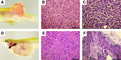
Tumors from clonal zebrafish can be transplanted into syngeneic recipient animals. Advanced stages of tumor development after intramuscular transplantation of moderately differentiated hepatocellular carcinoma zt34 (A, B) compared with normal liver (C). A spontaneous acinar cell carcinoma of the pancreas was capable of robust engraftment when intraperitoneally injected into clonal fish (D). Histological analysis showed that the tumor contained areas that were morphologically similar to normal pancreas (E, F, respectively). Scale bars (B, C, E, F) equal 50μm. |
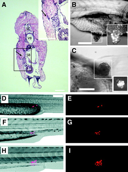
Human cancers can engraft into zebrafish embryos and larvae. Hematoxylin and eosin-stained cross section of a juvenile zebrafish that had been transplanted with MDA-435 adenocarcinoma cells. Cells were injected intraperitoneally and imaged at 5 days posttransplantation (Stoletov et al.56). Adenocarcinoma cells invaded the body wall (arrow, A). Anatomical structures include spinal cord (SC), vertebrae (VB), and swim bladder (SB). Transplanted metastatic WM-266-4 melanoma cells formed pigmented masses in the intestinal wall by 7 days postinjection (Haldi et al.60). Lateral view (B) and ventral view (C) of the same fish. Boxed area: pigmented tumor masses seen with brightfield illumination are also fluorescent-labelled with em-Di dye (bottom right). Red fluorescent protein–labeled human U251 glioblastoma cells can engraft into larval fish (D–I). Two U251 glioblastoma cells transplanted into a 2-day-old zebrafish embryo proliferate over time (Geiger et al.61): 2 days (D, E), 4 days (F, G), and 9 days posttransplantation (H, I). |