
Fig. 6
- ID
- ZDB-FIG-100114-9
- Publication
- Ignatius et al., 2009 - Zebrafish as a Model for Cancer Self-Renewal
- Other Figures
- All Figure Page
- Back to All Figure Page
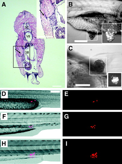
Human cancers can engraft into zebrafish embryos and larvae. Hematoxylin and eosin-stained cross section of a juvenile zebrafish that had been transplanted with MDA-435 adenocarcinoma cells. Cells were injected intraperitoneally and imaged at 5 days posttransplantation (Stoletov et al.56). Adenocarcinoma cells invaded the body wall (arrow, A). Anatomical structures include spinal cord (SC), vertebrae (VB), and swim bladder (SB). Transplanted metastatic WM-266-4 melanoma cells formed pigmented masses in the intestinal wall by 7 days postinjection (Haldi et al.60). Lateral view (B) and ventral view (C) of the same fish. Boxed area: pigmented tumor masses seen with brightfield illumination are also fluorescent-labelled with em-Di dye (bottom right). Red fluorescent protein–labeled human U251 glioblastoma cells can engraft into larval fish (D–I). Two U251 glioblastoma cells transplanted into a 2-day-old zebrafish embryo proliferate over time (Geiger et al.61): 2 days (D, E), 4 days (F, G), and 9 days posttransplantation (H, I). |