
- Title
-
Early regulation of brain aromatase (cyp19a1b) by estrogen receptors during zebrafish development
- Authors
- Mouriec, K., Lareyre, J.J., Tong, S.K., Le Page, Y., Vaillant, C., Pellegrini, E., Pakdel, F., Chung, B.C., Kah, O., and Anglade, I.
- Source
- Full text @ Dev. Dyn.
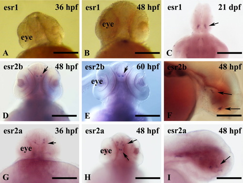
Distribution of esr1, esr2b, and esr2a mRNAs (arrows) in zebrafish embryos. A-I: Shown are whole-mount in situ hybridizations performed on embryos at 36 hours postfertilization (hpf; A,G), 48 hpf (B,D,F,H,I), 60 hpf (E), and 21 days postfertilization (adult brain, C). A, B, D, E, G, and H are frontal views; F and I are lateral views anterior to the left. D and E show a strong expression of esr2b mRNA in diencephalon at 48 and 60 hpf. F: Sagittal view reveals a weak signal in the hypothalamus. G-I: At 36 and 48 hpf, esr2a mRNA is present in restricted cell populations of the presumptive telencephalon, preoptic region, and hypothalamus of embryos. Scale bars = 200 μm in A,B,D,E, 50 μm in C, 100 μm in F,I, 250 μm in G,H. |

A-C: Expression of aromatase B messengers in the forebrain of zebrafish of 36 hours postfertilization (hpf) and 48 hpf. To detect messengers encoding aromatase B, embryos were treated with estradiol (E2) (10-8 M). A,B: Significant staining localized in preoptic area (POA) and hypothalamus (MBH). C: A transverse cryostat section in the preoptic area showing abundant cyp19a1b mRNA staining along the midline in radial glial cells and their processes in a 48 hpf embryo. D,E: Confocal images showing dorsal view of the head of cyp19a1b-GFP (green fluorescent protein) transgenic embryos demonstrating the fluorescence induced by a treatment with E2 for 26 hpf (B), while no signal is detected in the EtOH-treated controls (A). The GFP fluorescence is localized in the presumptive ventral diencephalon. Images correspond to the merge of a 20 images Z-stack. Bar = 100μm. F-I: Micrographs taken on horizontal sections showing that the cyp19a1b driven GFP expression (F) perfectly matches aromatase B immunoreactivity (G) in a 48 hpf larva treated with E2. The staining always corresponds to cells bordering the ventricles (F-H) and sending long cytoplasmic processes at the brain periphery (I), in this case in the anterior medial hypothalamus (MBH). Note that the nucleus of the cells is stained in by GFP but not by aromatase B (I). Also note that putative young neurons stained in blue by DAPI are aligned along the processes of the radial cells. Scale bar = 100 μm in A-C, 100 μm in D-H, 40 μm in I. |