
|
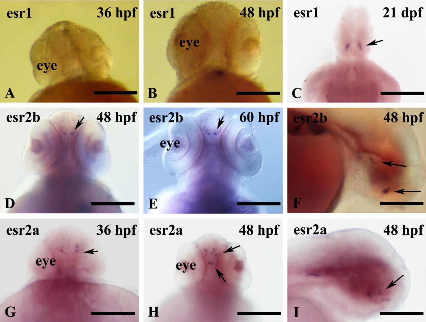
Fig. 2 Distribution of esr1, esr2b, and esr2a mRNAs (arrows) in zebrafish embryos. A-I: Shown are whole-mount in situ hybridizations performed on embryos at 36 hours postfertilization (hpf; A,G), 48 hpf (B,D,F,H,I), 60 hpf (E), and 21 days postfertilization (adult brain, C). A, B, D, E, G, and H are frontal views; F and I are lateral views anterior to the left. D and E show a strong expression of esr2b mRNA in diencephalon at 48 and 60 hpf. F: Sagittal view reveals a weak signal in the hypothalamus. G-I: At 36 and 48 hpf, esr2a mRNA is present in restricted cell populations of the presumptive telencephalon, preoptic region, and hypothalamus of embryos. Scale bars = 200 μm in A,B,D,E, 50 μm in C, 100 μm in F,I, 250 μm in G,H.