
|
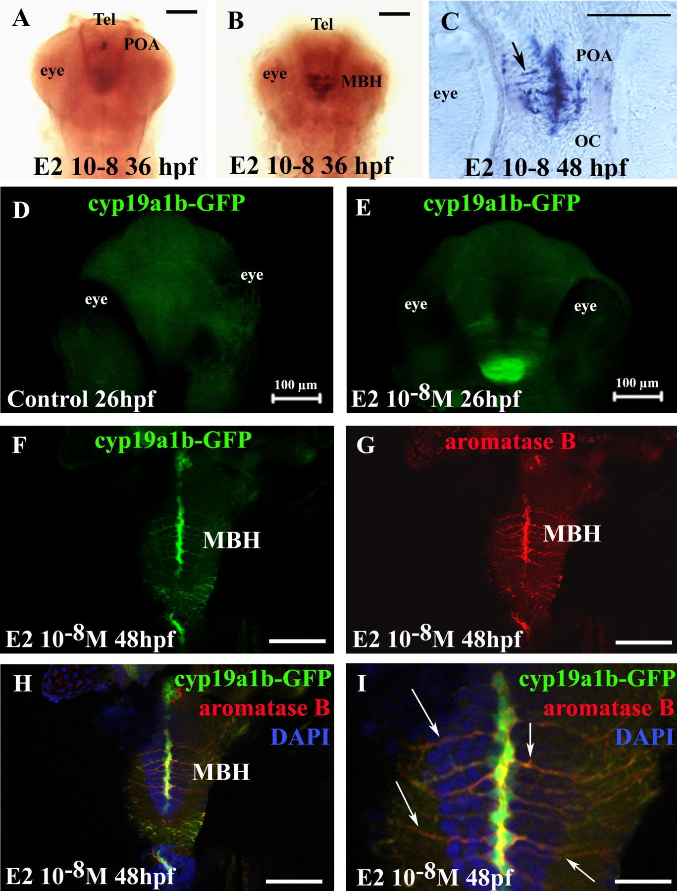
Fig. 4 A-C: Expression of aromatase B messengers in the forebrain of zebrafish of 36 hours postfertilization (hpf) and 48 hpf. To detect messengers encoding aromatase B, embryos were treated with estradiol (E2) (10-8 M). A,B: Significant staining localized in preoptic area (POA) and hypothalamus (MBH). C: A transverse cryostat section in the preoptic area showing abundant cyp19a1b mRNA staining along the midline in radial glial cells and their processes in a 48 hpf embryo. D,E: Confocal images showing dorsal view of the head of cyp19a1b-GFP (green fluorescent protein) transgenic embryos demonstrating the fluorescence induced by a treatment with E2 for 26 hpf (B), while no signal is detected in the EtOH-treated controls (A). The GFP fluorescence is localized in the presumptive ventral diencephalon. Images correspond to the merge of a 20 images Z-stack. Bar = 100μm. F-I: Micrographs taken on horizontal sections showing that the cyp19a1b driven GFP expression (F) perfectly matches aromatase B immunoreactivity (G) in a 48 hpf larva treated with E2. The staining always corresponds to cells bordering the ventricles (F-H) and sending long cytoplasmic processes at the brain periphery (I), in this case in the anterior medial hypothalamus (MBH). Note that the nucleus of the cells is stained in by GFP but not by aromatase B (I). Also note that putative young neurons stained in blue by DAPI are aligned along the processes of the radial cells. Scale bar = 100 μm in A-C, 100 μm in D-H, 40 μm in I.