
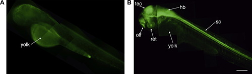
Fugu gap43 promoter drives expression in newly differentiating neurons of the embryo. 48 hpf zebrafish embryos: (A) Yolk autofluorescence of non-transgenic zebrafish; (B) tg(3.6fgap43:GFP)SA1 transgenic zebrafish displays transgene (GFP) expression throughout the developing nervous system: retina (ret), olfactory epithelia (olf), optic tectum (tec), hindbrain (hb), and spinal cord (sc). Scale bar = 200 μm. EXPRESSION / LABELING:
|
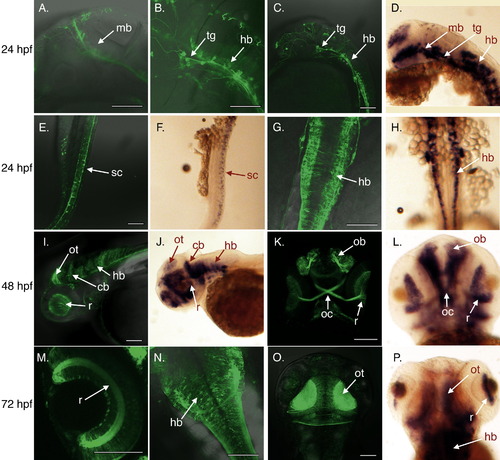
Fugu gap43 promoter mimics endogenous gap43 expression pattern in developing embryos. (A–C, E, and G) Neurons from 24 hpf tg(3.6fgap43:GFP)SA1 embryos expressing GFP in the midbrain (mb) and hind brain (hb), as well as neurons in the trigeminal ganglion (tg), and spinal cord (sc). Expression of endogenous gap43 determined by in situ hybridization show similar patterns of expression (D, F, H). 48 hpf: expression in the retina(r), cerebellum (cb), hindbrain (hb), olfactory bulb (ob), optic chiasm (oc), optic tectum (ot) observed for transgene (I, K) and endogenous gap43 (J, L). 72 hpf: expression in the retina (r), hindbrain (hb), optic tectum (ot) observed for transgene (M–O) and endogenous gap43 (P). (A–F, I, J) lateral views; (G, H, M–P) dorsal views; (K, L) ventral views. Scale bars = 100 μm. EXPRESSION / LABELING:
|
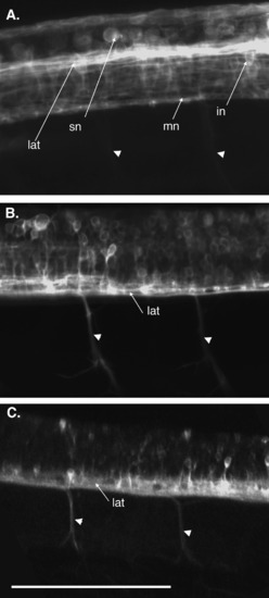
Transgene expression in spinal neurons is developmentally regulated. Lateral view of maximum projections of confocal stacks taken through tg(3.6fgap43:GFP)SA1 spinal cord at 24, 48, and 96 hpf. (A) Motorneurons (mn), interneurons (in), and sensory neurons (sn), as well as logitudinal axonal tracts (lat) are observed at 24 hpf. Signal from axons of motorneurons exiting the spinal cord is weak, but increases over time as the number of axons increase (arrowheads). (B) GFP-expressing cell bodies are observed throughout the dorso-ventral axis of the spinal cord, and axons of motorneurons (arrowheads) are observed exiting the spinal cord at 48 hpf. (C) By 96 hpf, expression in the cell bodies of spinal neurons is diminished, but longitudinal axonal tracts and axons of motorneurons exiting the spinal cord continue to express GFP. Arrowheads indicate motorneuron axons exiting the spinal cord. Scale bar = 100 μm. |
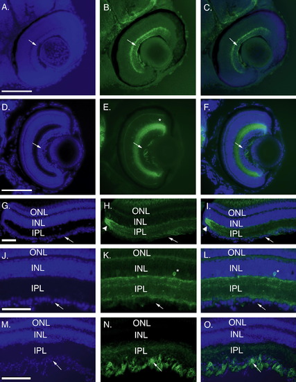
Transgene expression is reinitiated in retinal ganglion cells following optic nerve injury. Transgene expression in sectioned retinas from developing retinas (sagittal sections: A–C, 24 hpf; D–F, 4 dpf) and adult retina (cross sections: control, G-L; regenerating, M–O). DAPI staining (A, D, G, J, M), GFP fluorescence (B, E, H, K, N), and merged images (C, F, I, L, O) are shown. Transgene expression is localized to the differentiated ganglion cell layer (GCL, arrows) at 48 hpf (B, C). By 96 hpf all layers of the retina have formed and expression is localized to the inner plexiform layer (IPL), with a few cells in the inner nuclear layer (INL, asterisk in E) and absent from the GCL (arrows; E, F). In control adult retina (G–L) GFP expression remains localized to the IPL (K, L) and a few cells in the INL (asterisk) and absent from the GCL (arrows). There is also bright expression at the retinal margin (arrowheads H, I). High levels of GFP expression are observed in the GCL 7 days after optic nerve crush (arrows N, O). ONL, outer plexiform layer. Scale bars = 100 μm. |
Reprinted from Gene expression patterns : GEP, 8(6), Udvadia, A.J., 3.6kb Genomic sequence from Takifugu capable of promoting axon growth-associated gene expression in developing and regenerating zebrafish neurons, 382-388, Copyright (2008) with permission from Elsevier. Full text @ Gene Expr. Patterns