
|
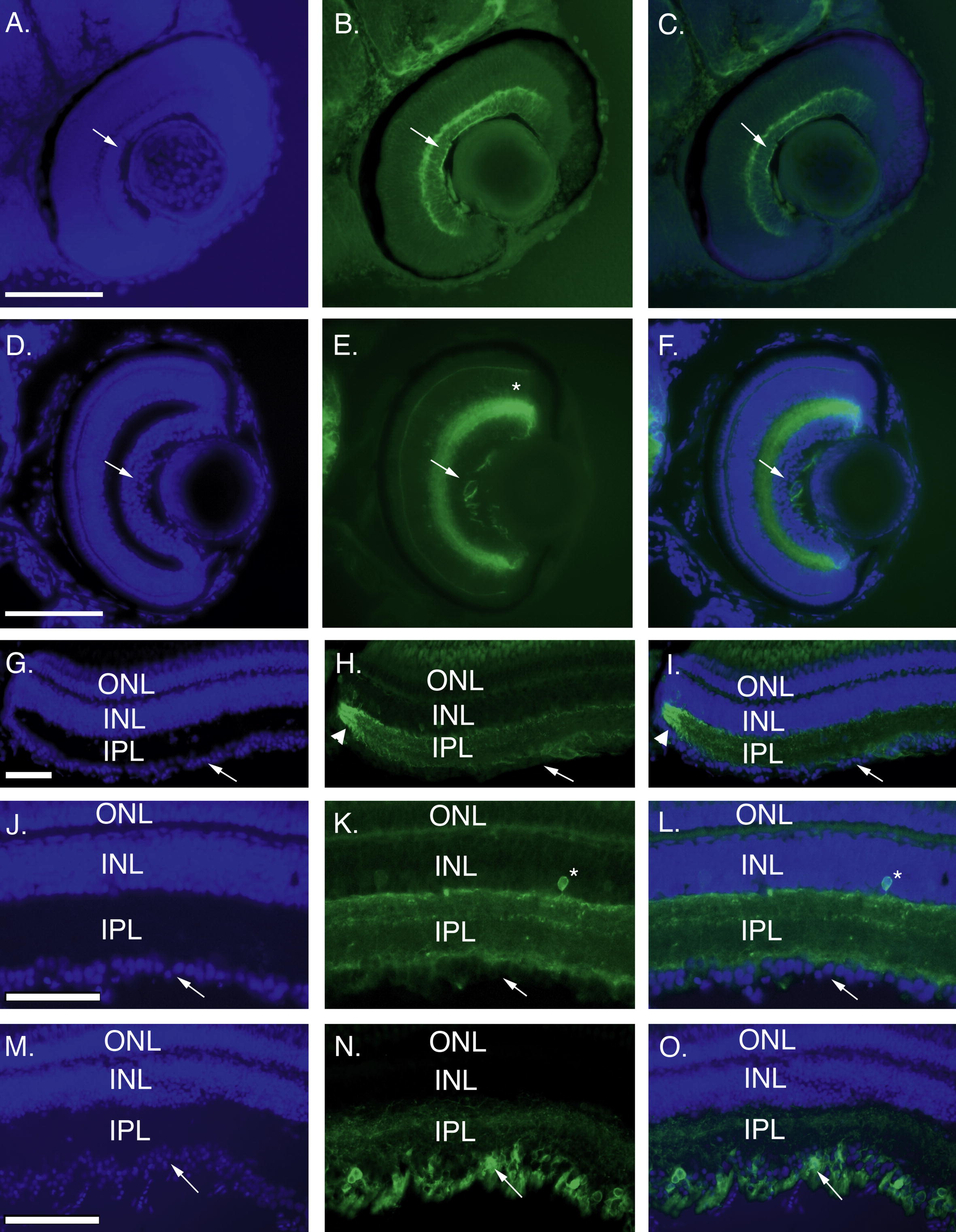
Fig. 6 Transgene expression is reinitiated in retinal ganglion cells following optic nerve injury. Transgene expression in sectioned retinas from developing retinas (sagittal sections: A–C, 24 hpf; D–F, 4 dpf) and adult retina (cross sections: control, G-L; regenerating, M–O). DAPI staining (A, D, G, J, M), GFP fluorescence (B, E, H, K, N), and merged images (C, F, I, L, O) are shown. Transgene expression is localized to the differentiated ganglion cell layer (GCL, arrows) at 48 hpf (B, C). By 96 hpf all layers of the retina have formed and expression is localized to the inner plexiform layer (IPL), with a few cells in the inner nuclear layer (INL, asterisk in E) and absent from the GCL (arrows; E, F). In control adult retina (G–L) GFP expression remains localized to the IPL (K, L) and a few cells in the INL (asterisk) and absent from the GCL (arrows). There is also bright expression at the retinal margin (arrowheads H, I). High levels of GFP expression are observed in the GCL 7 days after optic nerve crush (arrows N, O). ONL, outer plexiform layer. Scale bars = 100 μm.
Reprinted from Gene expression patterns : GEP, 8(6), Udvadia, A.J., 3.6kb Genomic sequence from Takifugu capable of promoting axon growth-associated gene expression in developing and regenerating zebrafish neurons, 382-388, Copyright (2008) with permission from Elsevier. Full text @ Gene Expr. Patterns