
- Title
-
The Tg(ccnb1:EGFP) transgenic zebrafish line labels proliferating cells during retinal development and regeneration
- Authors
- Kassen, S.C., Thummel, R., Burket, C.T., Campochiaro, L.A., Harding, M.J., and Hyde, D.R.
- Source
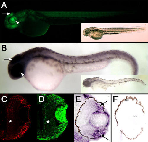
Expression of the cyclin B1:EGFP transgene at 48 hpf. A: A fluorescent image of a Tg(ccnb1:EGFP)nt18 embryo at 48 hpf is displayed. EGFP was expressed throughout the body of the fish with the most intense expression in the brain (arrow) and eye field (arrowhead). A bright-field image of the same embryo is shown in the inset. B: Whole-mount in situ hybridization revealed cyclin B1 mRNA expression throughout the body with the greatest staining in the head (arrow) and eye (arrowhead). A zebrafish labeled with the cyclin B1 sense RNA probe is shown in the inset. Retinal sections at 48 hpf reveal strong PCNA expression in cells in the retinal margin (C), which is similar to EGFP expression (D). EGFP and PCNA expression is decreased in the central retina (*). Retinal sections labeled with the cyclin B1 antisense RNA probe demonstrated a similar high level of expression near the retinal margin (E, arrows), while the control sense RNA probe did not label any retinal cells. F: No signal was observed with the sense RNA probe. Abbreviations: RPE represents retinal pigmented epithelium, and GCL represents ganglion cell layer. EXPRESSION / LABELING:
|
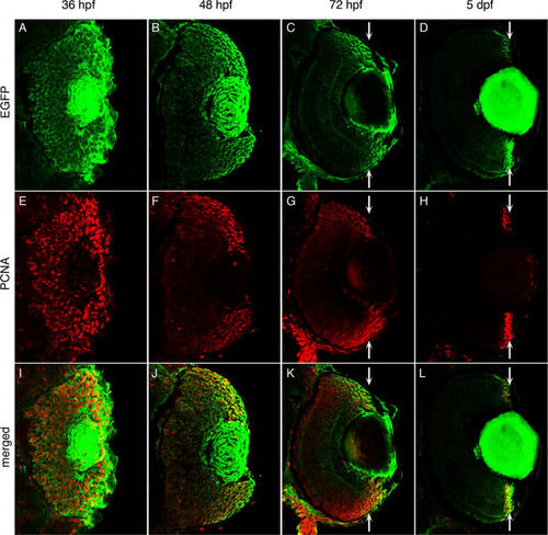
Enhanced green fluorescent protein expression during retinal development in Tg(ccnb1:EGFP)nt18 zebrafish. Enhanced green fluorescent protein (EGFP; A-D), proliferating cell nuclear antigen (PCNA; E-H), and merged expression patterns (I-L) are shown at 36, 48, 72 hpf, and 5 dpf of retinal development. At 36 hpf, PCNA and EGFP expression are almost ubiquitous throughout the retina (A, E, I). At 48 hpf, EGFP and PCNA expression is limited in the central retina, but persists near the retinal margin (B, F, J). At 72 hpf, EGFP and PCNA expression are largely absent in the central retina and become further focused in the cells near the margin (C, G, K; arrows). By 5 dpf, EGFP and PCNA expression are restricted to the retinal margin (D, H, L; arrows). At all time points, EGFP expression but not PCNA persists in the lens. |
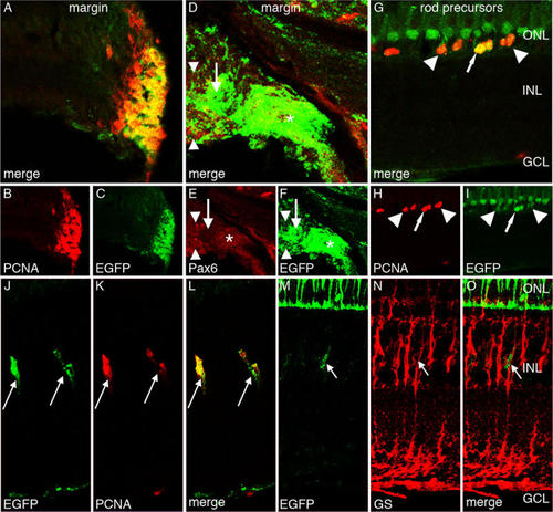
Enhanced green fluorescent protein expression in the Tg(ccnb1:EGFP)nt18 adult zebrafish retina. Enhanced green fluorescent protein (EGFP; C, F, I, J, M), proliferating cell nuclear antigen (PCNA; B, H, K), Pax6 (E), glutamine synthetase (GS; N), and merged expression (A, D, G, L, O) are shown. EGFP and PCNA are strongly expressed in the cells of the adult retinal circumferential marginal zone (CMZ; A-C). EGFP expression in the peripheral CMZ co-labels with Pax6 (*), but does not co-label near the distal CMZ (D-F, arrow). The Pax6-positive EGFP-negative signal adjacent to the CMZ (arrowheads) corresponds to the newly differentiated amacrine and ganglion cells. EGFP is also expressed in PCNA-positive rod precursor cells, which reside in the outer nuclear layer (G-I). Some of these rod precursor cells express high levels of EGFP (arrows), while others express lower EGFP levels (arrowhead). EGFP is also expressed in a row of nonproliferative PCNA-negative short single cones (G, I). Some Müller glia in the adult undamaged retina slowly divide and give rise to retinal progenitor cells. PCNA marks these proliferating Müller glial cells, which also coexpress EGFP (J-L, arrows). EGFP-positive cells in the INL also co-label with glutamine synthetase (M-O, arrow). This demonstrates that proliferating EGFP-positive cells co-label with Müller glia. Abbreviations: ONL represents outer nuclear layer; INL represents inner nuclear layer; and GCL represents ganglion cell layer. EXPRESSION / LABELING:
|
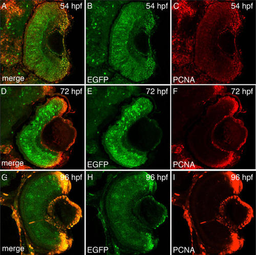
Enhanced green fluorescent protein expression during retinal development in Tg(1016α1tubulin:EGFP) zebrafish. Enhanced green fluorescent protein (EGFP; B, E, H), proliferating cell nuclear antigen (PCNA; C, F, I), and merged expression patterns (A, D, G) are shown at 54 hpf, 72 hpf, and 96 hpf of retinal development. At 54 hpf, EGFP is expressed throughout the entire retina, while PCNA expression is lower in the central retina relative to the retinal margin (A-C). At 72 hpf, EGFP continues to be expressed throughout the central retina, while PCNA expression is largely absent in the central retina and highly expressed in the cells near the margin (D-F). By 96 hpf, EGFP expression decreases in the central retina but remains in the margin, while PCNA expression remains restricted to the retinal margin (G-I). |
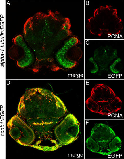
Enhanced green fluorescent protein and proliferating cell nuclear antigen expression in Tg(1016α1tubulin:EGFP) and Tg(ccnb1:EGFP)nt18 72 hpf larva head sections. Enhanced green fluorescent protein (EGFP; C, F), proliferating cell nuclear antigen (PCNA; B, E), and merged expression patterns (A, D) are shown in 72 hpf larval head sections. In the Tg(1016α1tubulin:EGFP) zebrafish, EGFP was predominantly expressed throughout the retina, while PCNA was expressed in the retinal margin, mandibular tissue, the dorsal epithelium, and tectum (A-C). In the Tg(ccnb1:EGFP)nt18 zebrafish, EGFP was restricted to the retinal margins similar to PCNA, but EGFP was also expressed in other head tissues that coexpressed PCNA (D-F). EXPRESSION / LABELING:
|