FIGURE SUMMARY
Title

Subfunctionalization of Duplicated Zebrafish pax6 Genes by cis-Regulatory Divergence

Authors
Kleinjan, D.A., Bancewicz, R.M., Gautier, P., Dahm, R., Schonthaler, H.B., Damante, G., Seawright, A., Hever, A.M., Yeyati, P.L., van Heyningen, V., and Coutinho, P.
Source
Full text @ PLoS Genet.

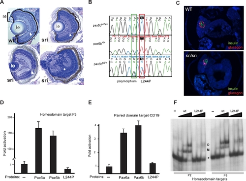
Characterisation of the sunrise (sri) Mutant
(A) Sections through wild type and homozygous sri larval eyes at 5 dpf, showing variable severity from mildly reduced lens size to almost complete absence of lens and retinal malformation (*). le, lens; re, neural retina and retinal pigment epithelium (black layer); arrowhead, optic nerve.
(B) Sequence traces from pax6b homeodomain revealing the T to C mutation resulting in the L244P mutation. The G to A third position change two bases earlier is a polymorphism between the WIK and Tü strains.
(C) Expression of known pax6 targets, glucagon and insulin, in pancreas of wild type (wt) 5 dpf fish. Expression is maintained in sri despite the loss-of-function homeodomain missense mutation in Pax6b, the sole pax6 gene expressed in the pancreas.
(D–F) Functional analysis of the Pax6bsri (L244P) protein in comparison with wild type Pax6b and Pax6a, using luciferase reporter assays in HeLa cells and EMSA.
(D) Pax6a and Pax6b proteins drive luciferase expression at comparable levels under the control of the P3 homeodomain promoter; the L244P mutant of Pax6b protein fails to activate P3.
(E) Pax6a and Pax6b proteins drive comparable luciferase expression levels through the CD19 paired domain target promoter; the L244P mutant of Pax6b protein has significantly reduced activity.
(F) Gel-retardation assay to analyse the binding capacity to homeodomain target binding sites P2 and P3 demonstrates reduced affinity of the L244P mutant protein compared to wild type (wt) Pax6b.
D, dimeric protein-bound; M, monomeric protein-bound; and F, free labeled target oligonucleotide. Protein concentrations used 0.5, 1.5, and 4.5 μM (lanes from left to right) with 5 nM oligonucleotides.

PHENOTYPE:
Fish:
Observed In:
Stage: Day 5

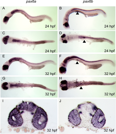
pax6a and pax6b Expression Analysis by Wholemount RNA in situ Hybridisation during Early Zebrafish Development
pax6a expression is shown in (A) and (C) at 24 hpf; (A) lateral view showing expression in eye, telencephalon, diencephalon, hind brain, and neural tube (C) dorsal view; (E) and (G) lateral and dorsal view at 32 hpf, showing continuing pattern of expression, (I) Vibratome coronal section of wholemount stained embryo expressing pax6a in telencephalon (arrowed), diencephalon, retina and lens. (B) pax6b expression is seen in lateral view showing eye and optic tectum at 24 hpf, pancreas expression is highlighted using black triangles; (D) dorsal view at 24 hpf. (F) and (H) lateral and dorsal. Increasing hindbrain and neural tube expression of pax6b is revealed by 32 hpf embryos. (J) predominantly eye expression of pa6b is seen in coronal section at 32 hpf.

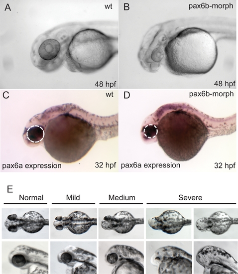
Analysis of pax6b Function in Zebrafish, with the Aim of Validating the Effect of the pax6b L244P Missense Mutation in sri Fish
(A–D) Phenotype elicited using pax6b morpholinos: (A) Uninjected wild type fish at 48 hpf; fish injected with the same amount of a control scrambled morpholino also produced no effect.
(B) Substantially reduced eye size and generally delayed development are observed by 48 hpf in response to injection of either of the two pax6b morpholinos.
(C and D) At 32 hpf reduction in eye size is observed (white dotted outlines) in otherwise normal looking embryos.
(E) The effects of injecting capped mRNA at levels that generally rescue the phenotype illustrating the spectrum of phenotypes to bilateral anophthalmia, observed when pax6b capped mRNA is injected at levels generally used for phenotype rescue experiments in zebrafish.

EXPRESSION / LABELING:
Gene:
Fish:
Knockdown Reagents:
Anatomical Terms:
Stage: Prim-15
PHENOTYPE:
Fish:
Knockdown Reagents:
Observed In:
Stage Range: Prim-15 to Long-pec

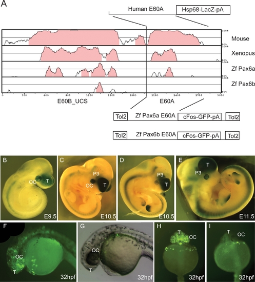
Enhancer Capacity of the E60A Conserved Element Assessed in Transgenic Mice and Zebrafish.
(A) Vista plot of the region around the highly conserved region E60+, located 47 kb downstream from the PAX6 P1 promoter. Human sequence is used as the base for pairwise comparison with pax6 loci from mouse, Xenopus tropicalis, and zebrafish (pax6a and pax6b). Fragment E60B_UCS contains an ultraconserved element that will be described elsewhere and is used here as an anchor point to allow localisation of the conserved/non-conserved E60A element from zebrafish pax6a and pax6b. A fragment containing the human E60A element was cloned into an hsp68-LacZ reporter construct to generate transient transgenic embryos. The equivalent regions from the zebrafish pax6a and pax6b loci were cloned into a Tol2 transposon based vector containing a cFos minimal promoter GFP reporter cassette [53] and used to produce transgenic zebrafish.
(B–E) Reporter expression of the human E60A construct was observed in optic cup, telencephalon, and prosomere P3 region of the diencephalon in embryos of E9.5 (B); E10.5 (C and D); and E11.5 (E). Expression (ectopic) in the limbs is ascribed to the genomic integration site of the construct.
(F) A similar GFP reporter expression pattern is seen in transgenic zebrafish embryos with the E60A region from the pax6a locus, with GFP expression observed in the optic cup and forebrain.
(G) No reporter expression is seen with the construct containing the E60A region from locus pax6b.
(H) Frontal view of a transgenic zebrafish with the pax6a E60A element, showing expression in telencephalon and optic cup.
(I) Frontal view of a pax6b E60A transgenic fish showing a lack of specific expression. T, telencephalon; OC, optic cup; P3, prosomere P3 of the diencephalon.

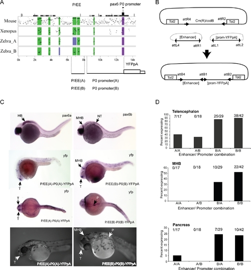
Transgenic Analysis of the Zebrafish P/EE Enhancers from the pax6a and pax6b Loci in Combination with the pax6 P0 Promoters
(A) PIP plot showing the conservation levels in the Pax6 upstream regions of mouse, Xenopus tropicalis, and the two zebrafish co-orthologues using the human gene as base sequence. The position of the pancreas/ectodermal (P/EE) and the P0 promoter regions used in the construction of the reporter transgenes are shown.
(B) Schematic representation of the Tol2-2way system to create combinatorial constructs for Tol2 mediated zebrafish transgenesis.
(C) Expression analysis of pax6a and pax6b shown by RNA in situ analysis with alkaline phosphatase staining at 28 hpf (top). YFP expression in P/EE(A)-P0(A)-YFPpA (left) and P/EE(B)-P0(B)-YFPpA (right) transgenic fish at 28 hpf shown, using in situ analysis for YFP mRNA. 56 hpf fluorescence images of YFP expression in transgenic lines for the same two constructs are shown on the bottom row.
(D) Frequency of expression in different sites in transient transgenic zebrafish with the four combinations of P/EE enhancer and P0 promoter from the two pax6 loci. T, telencephalon; MHB, midbrain hindbrain boundary; HB, hindbrain; P, pancreas; NT, neural tube.

EXPRESSION / LABELING:
Genes:
Fish:
Anatomical Terms:
Stage: Prim-5

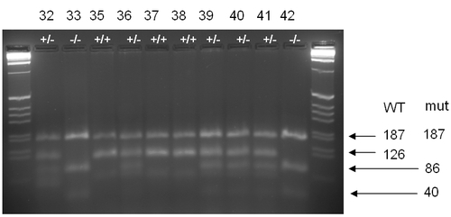
Genotyping of sri mutant and wild type larvae
DdeI restriction digest of fragment spanning the sri mutation site, showing the genotyping of fish from sri/+ x sri/+ parental cross. Genotyping was carried out on the 5 dpf larvae that were analysed for the pancreatic phenotype in Figure 1 of the paper.
The ratio of WT : heterozygous : homozygous mutant genotypes did not deviate significantly from the expected frequency of 1:2:1, with a Chi square (Σ2) value of 1.390, showing that the deviation observed is not significant, with a p value of 0.499. (observed ratios 7:23:11 at 5 dpf)

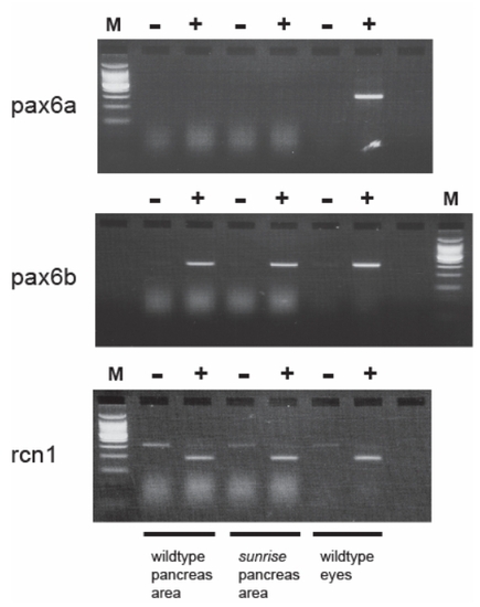
RT-PCR Analysis Shows That Only pax6b Is Expressed in the Pancreas of Both Wild Type and sri/sri Homozygous Adult Fish. In Contrast, Both pax6a and pax6b Are Expressed in Adult Eye.
Only pax6b is expressed in adult wild type and sri/sri pancreas, while both pax6a and pax6b are expressed from adult eyes isolated from the same individuals

EXPRESSION / LABELING:
Genes:
Fish:
Anatomical Terms:
Stage: Adult

Unillustrated author statements

EXPRESSION / LABELING:
Gene:
Fish:
Anatomical Term:
Stage: Adult
Acknowledgments
This image is the copyrighted work of the attributed author or publisher, and ZFIN has permission only to display this image to its users. Additional permissions should be obtained from the applicable author or publisher of the image. Full text @ PLoS Genet.