Transgenic Analysis of the Zebrafish P/EE Enhancers from the pax6a and pax6b Loci in Combination with the pax6 P0 Promoters
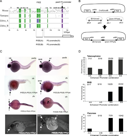
(A) PIP plot showing the conservation levels in the Pax6 upstream regions of mouse, Xenopus tropicalis, and the two zebrafish co-orthologues using the human gene as base sequence. The position of the pancreas/ectodermal (P/EE) and the P0 promoter regions used in the construction of the reporter transgenes are shown.
(B) Schematic representation of the Tol2-2way system to create combinatorial constructs for Tol2 mediated zebrafish transgenesis.
(C) Expression analysis of pax6a and pax6b shown by RNA in situ analysis with alkaline phosphatase staining at 28 hpf (top). YFP expression in P/EE(A)-P0(A)-YFPpA (left) and P/EE(B)-P0(B)-YFPpA (right) transgenic fish at 28 hpf shown, using in situ analysis for YFP mRNA. 56 hpf fluorescence images of YFP expression in transgenic lines for the same two constructs are shown on the bottom row.
(D) Frequency of expression in different sites in transient transgenic zebrafish with the four combinations of P/EE enhancer and P0 promoter from the two pax6 loci. T, telencephalon; MHB, midbrain hindbrain boundary; HB, hindbrain; P, pancreas; NT, neural tube.
|
