
- Title
-
Gene expression analysis on sections of zebrafish regenerating fins reveals limitations in the whole-mount in situ hybridization method
- Authors
- Smith, A., Zhang, J., Guay, D., Quint, E., Johnson, A., and Akimenko, M.A.
- Source
- Full text @ Dev. Dyn.
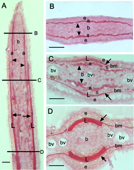
Picrosirius red staining of collagen on sections of 4-day regenerates. Longitudinal (A) and transverse (B-D) cryo-sections of a 4 dpa regenerated fin ray that has been stained with picrosirius red, which labels collagenous structures, including the lepidotrichia (arrows), the actinotrichia (arrowheads) and the basement membrane. The horizontal lines in A indicate the approximate positions of the transverse sections presented in B-D along the proximal-distal axis of the regenerate. The level of amputation of the fin is not shown in A. Note the presence of the tip of the actinotrichia in the distal part of the regenerate (B). More proximally, both actinotrichia of increased diameter and the lepidotrichia bone matrix are visible (C), while in the proximal part of the regenerate only thick lepidotrichia are present (D). L, lepidotrichia; a, actinotrichia; b, blastema; e, epidermis; bm, basement membrane; bv, blood vessels; s, scleroblasts. Scale bars = 25 μm (A-D). |

Comparison of the expression pattern of two bone markers, col10a1 (A, C, D, C′, D′) and Zns-5 (B, E, F, E′, F′), within the fin regenerate following two different in situ hybridization and immunostaining techniques. Whole-mount in situ hybridization (ISH) with col10a1 RNA antisense probe (A) and immunostaining with Zns-5 antibody (B) on 4-dpa regenerates. Arrows in A and B indicate the level of amputation. C, C′, E, E′: Longitudinal cryo-sections of the whole-mount 4-dpa fin regenerates shown in A and B. D, D′, F, F′: ISH of col10a1 probe (D, D′) and immunostaining using Zns-5 antibody (F, F′) performed on longitudinal cryo-sections of 4-dpa regenerates. C: col10a1 expression on sections following ISH is found in the basal layer of the epidermis and also in the scleroblast cells lining the outer lepidotrichia. D: In addition, when ISH is performed on cryo-sections, col10a1 expression is also detected in the scleroblasts lining the inner side of the regenerating lepidotrichia. C′, D′: Higher magnification of the area indicated by the dashed box in C and D, respectively. Black arrowheads in C′ and D′ indicate expression in scleroblasts. Note that the inner scleroblasts (right arrowhead in D′) are only detected when an ISH is performed on cryo-sections. The asterisk in C′ indicates the absence of detected expression in the inner scleroblasts when sections are done following a whole-mount ISH. E: On sections obtained after immunostaining on whole-mount samples, the Zns-5 antibody is staining all the outer scleroblasts along the entire length of the regenerate and the inner scleroblasts located in the distal part of the regenerate but not the more proximal one, which are only detected following immunostaining on cryo-sections (F). E′, F′: Higher magnification of the area indicated by the dashed box in E and F, respectively. Black arrowheads in E′ and F′ indicate expression in scleroblasts. Note that staining in the inner scleroblasts (right arrowhead in F′) is only detected when the procedure is performed directly on cryo-sections. The asterisk in E′ indicates the absence of detection in the inner scleroblasts when sections are done following immunodetection on a whole-mount sample. L, lepidotrichia; b, blastema; ble, basal layer of epidermis; s, scleroblasts. Scale bars = 100 μm (A,B), 50 μm (C,D), 25 μm (C′, D′, E, E′, F, F′). EXPRESSION / LABELING:
|

Comparison of the expression of msxb and msxd genes in 4-day fin regenerates using the two ISH methods. A,B: Whole-mount ISH showing expression of msxb and msxd in the distal region of the fin regenerate at 4 dpa. msxd is also expressed in the distal part of the interray tissue. C: Upon sectioning of the whole-mount fins, msxb expression is seen in the distal blastema. D: ISH on sections reveals a broader expression domain for msxb encompassing the entire blastema as well as in newly differentiating scleroblasts. E: Sections of whole-mount fins hybridized with the msxd probe shows expression in the distal epidermis, in the distal blastema, and, on some sections, in the basal layer of the epidermis adjacent to the differentiating scleroblasts. F: msxd expression following ISH on sections is similar to that seen following whole-mount ISH. Arrowheads in E and F indicate the basal epidermal cells close to the differentiating scleroblasts expressing msxdF: msxd expression following ISH on sections is similar to that seen following whole-mount ISH. Arrowheads in E and F iE. Arrows in A and B indicate the level of amputation. L, lepidotrichia; b, blastema; ble, basal lay EXPRESSION / LABELING:
|
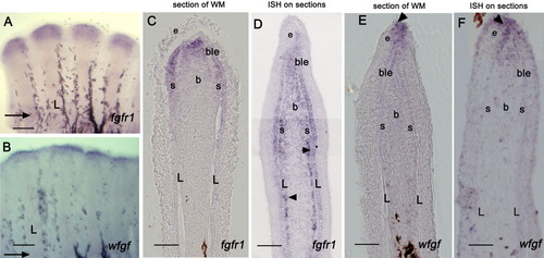
Comparison of the expression of fgfr1 and wfgf genes in 4-day fin regenerates using the two ISH methods. A, B: Whole-mount ISH showing expression of fgfr1 and wfgf in the distal region of the fin regenerate at 4 dpa. C: Upon sectioning of the whole-mount fins, fgfr1 expression is seen in the distal blastema, in the basal layer of the epidermis, and in a few cells of the newly differentiating scleroblasts. D: ISH on sections reveals a broader expression domain for fgfr1: transcripts are observed in the blastema midway through the proximal-distal axis. It is observed, not only in newly differentiating scleroblasts, but also in differentiated scleroblasts in more proximal regions lining the inner side of the lepidotrichia (arrowheads in D indicate scleroblasts). E: Sections of whole-mount fins hybridized with the wfgf probe shows that wfgf expression is confined to the epidermis in the most distal tip of the regenerate (arrowhead). F: wfgf expression following ISH on sections is similar to that seen following whole-mount ISH (arrowhead). Arrows in A and B indicate the level of amputation. L, lepidotrichia; b, blastema; ble, basal layer of epidermis; e, epidermis; s, scleroblasts. Scale bars = 100 μm (A,B), 50 μm (C-F). EXPRESSION / LABELING:
|
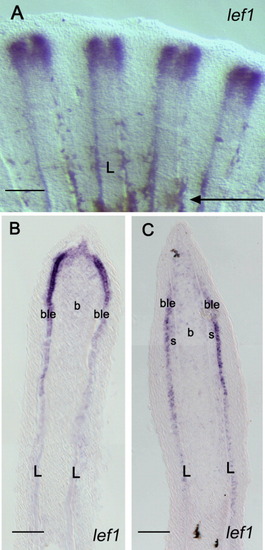
Comparison of the expression of lef1 in a 4-day fin regenerate using the two ISH methods. A: Whole-mount ISH with a lef1 probe on 4-dpa regenerates shows expression splitting into two domains in the distal part of each fin ray indicating an imminent bifurcation event. B: Upon cryo-sectioning of the fin regenerate shown in A, lef1 expression is observed in the basal layer of the epidermis and in the distal blastema. C: A similar expression pattern is observed when an ISH is performed on cryo-sections of 4-dpa regenerates. The arrow in A indicates the level of amputation. L, lepidotrichia; b, blastema; ble, basal layer of epidermis; s, scleroblasts. Scale bars = 100 mu;m (A), 50 μm (B,C). EXPRESSION / LABELING:
|