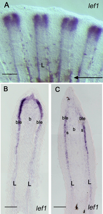
Fig. 5 Comparison of the expression of lef1 in a 4-day fin regenerate using the two ISH methods. A: Whole-mount ISH with a lef1 probe on 4-dpa regenerates shows expression splitting into two domains in the distal part of each fin ray indicating an imminent bifurcation event. B: Upon cryo-sectioning of the fin regenerate shown in A, lef1 expression is observed in the basal layer of the epidermis and in the distal blastema. C: A similar expression pattern is observed when an ISH is performed on cryo-sections of 4-dpa regenerates. The arrow in A indicates the level of amputation. L, lepidotrichia; b, blastema; ble, basal layer of epidermis; s, scleroblasts. Scale bars = 100 mu;m (A), 50 μm (B,C).
Image
Figure Caption
Figure Data
Acknowledgments
This image is the copyrighted work of the attributed author or publisher, and
ZFIN has permission only to display this image to its users.
Additional permissions should be obtained from the applicable author or publisher of the image.
Full text @ Dev. Dyn.

