
- Title
-
Mechanisms controlling Pax6 isoform expression in the retina have been conserved between teleosts and mammals
- Authors
- Lakowski, J., Majumder, A., and Lauderdale, J.D.
- Source
- Full text @ Dev. Biol.

Structure and transcript analysis of the zebrafish Pax6a dual-reporter BAC transgene. (A) Schematic representation of zebrafish BAC DKEY-46C10 showing the location of the Pax6a transcript unit within the BAC. Black boxes denote exons, and bent arrows denote promoters P0, P1, and Palpha. (B) The zebrafish Pax6a BAC was modified by insertion of an enhanced green fluorescent protein reporter cassette (EGFP pA) in-frame with the evolutionarily conserved ATG in exon 4. A Discosoma red fluorescent protein reporter cassette (DsRed-pA) was inserted in-frame into the conserved ATG in exon 8. (C) Whereas transcripts that initiate from the P0 and P1 promoters encode for EGFP, Palpha-initiated transcripts encode for DsRed. (D) Analysis of transcripts from the BAC transgene. RNA was prepared from 3-day old embryos transiently transgenic for the dual-reporter BAC and analyzed by RT-PCR. Transcripts with exon 1 (e1) or exon 2 (e2) also included EGFP, but not DsRed. Transcripts with exons alpha, 5 (e5), 6 (e6), or 7 (e7) also included DsRed. No P1- or P0-initiated transcripts contained DsRed. |

EGFP is expressed in a Pax6a-like pattern in developing zebrafish. (A, C, E) Pax6a expression visualized by whole-mount mRNA in situ hybridization. (B, D, F) Transgene expression visualized by EGFP fluorescence. EGFP was expressed by cells within domains of Pax6a expression. These results suggest that BAC DKEYP-46C10 is sufficient to recapitulate the Pax6a expression pattern in all regions of the developing zebrafish. |

DsRed expression is restricted to the developing zebrafish eye. (A) Live zebrafish embryo at 3-days post fertilization (dpf) transiently transgenic for the dual-reporter Pax6a BAC. In this embryo, cells in the eye, diencephalon, hindbrain, and spinal cord express EGFP (B), but only cells in the eye express DsRed (C). DsRed expression in the eye was typically first detected between 2.0 and 2.5 dpf. |

Amacrine cells in the developing zebrafish retina express DsRed. Sections cut through the eye of transiently transgenic embryos at different stages of development. (A) At 28 hpf EGFP, but not DsRed, is expressed by retinal progenitor cells (arrowheads). EGFP-expressing cells are also visible in the diencephalon (di) and hindbrain (*). Inset: lateral view of a 28 hpf embryo showing the plane of section. Rostral is left and dorsal is up. (B) Starting at the time of amacrine cell differentiation (50 hpf), a subset of EGFP-expressing cells begin expressing DsRed (arrows). (C, D) Whereas differentiating cells in the ONL, INL, and GCL expressed EGFP, only amacrine cells appeared to express DsRed. EGFP expression in retinal ganglion cell axons was visible within the optic nerve by 72 hpf (on). Cell nuclei in D are labeled (blue) with Hoechst. (E–H) Sections cut through the eye from four different transiently transgenic embryos that were allowed to develop to 72 hpf. (E, F) DsRed is expressed by displaced amacrines (arrows), which have cell bodies in the GCL. (G) DsRed-expressing cells (red) coexpress Pax6 (immunostained in green). (H) DsRed-expressing cells (red) coexpress syntaxin (immunostained in blue), which is expressed in the dendritic processes and perikarya of all amacrine cells. hb, hindbrain; ln, lens; nr, neural retina. |
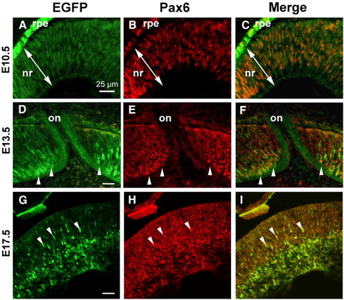
Zebrafish Pax6a P0/P1 promoters are active in Pax6-expressing cells in the developing mouse retina. Sections cut through the eyes of transgenic mouse embryos at E10.5 (A–C), E13.5 (D–F), or E17.5 (G–I). Both EGFP (A, D, G) and endogenous Pax6 (B, E, H) were visualized by indirect immunofluorescence (EGFP, green; Pax6, red). (C, F, I) Merged images. EGFP and Pax6 are coexpressed in cells in the neural retina (nr). At E10.5, EGFP expression replicated mouse Pax6. At both E13.5 (D–F) and E17.5 (G–I), EGFP was more strongly expressed in putative differentiating neurons (arrowheads) than in neuroepithelial cells. At E13.5 EGFP expression in retinal ganglion cell axons is visible within the optic nerve (on). Arrowheads in D–F denote ganglion cells with axonal projections into the developing optic nerve. Scale bars = 25 μm; bar in panel A applies to panels B–C; bar in panel D applies to panels E–I. |

Zebrafish Pax6a P0/P1 promoters are active in Pax6-expressing cells in the adult mouse retina. (A–B) EGFP from the zebrafish Pax6a BAC transgene is expressed by cells in the inner nuclear layer (INL) and ganglion cell layer (GCL). (A) EGFP+ radial processes also span the outer nuclear layer (ONL) and end at the outer limiting membrane (arrow). This suggests that Müller glia express the zebrafish Pax6a BAC transgene. (B–D) Direct comparison of EGFP and Pax6 expression in the adult mouse retina revealed that most, if not all, Pax6+ cells coexpressed EGFP; however, a few EGFP+ cells did not express Pax6 (arrowheads). The ectopic expression in these cells is most likely due to either species differences in expression or misregulation of the zebrafish Pax6a BAC transgene in mice. Scale bar in panel B applies to panels C–D. |
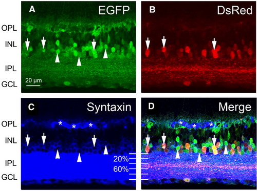
Paired-containing and paired-less transcripts from the zebrafish Pax6a BAC transgene are differentially expressed by amacrine cells in the adult mouse retina. (A, B, D) A subset of EGFP-expressing cells in the adult mouse retina coexpress DsRed. Colabeling with syntaxin (C, D) reveals three populations of amacrine cells: syntaxin+/EGFP-/DsRed- (blue cells in panel D), syntaxin+/EGFP+/DsRed- (arrowheads) and syntaxin+/EGFP+/DsRed+ (arrows). DsRed-expressing amacrine cells tend to project neurites to the middle strata of the IPL. Strata divisions were determined based on work by Masland and co-workers ([Masland, 2001] and [Masland, 2004]). Asterisks denote non-specific staining from a secondary antibody needed for these triple-label experiments. Scale bar in panel A applies to all panels. |
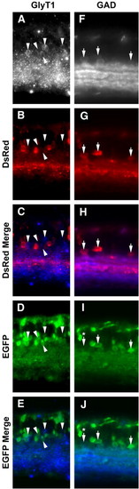
Zebrafish Pax6a paired-less transcripts are expressed in GABAergic, but not glycinergic, amacrine cells in the adult mouse retina. (A–E) Comparison of glycine transporter 1 (GlyT1), DsRed, and EGFP expression by coimmunostaining reveals that GlyT1+ cells express EGFP but not DsRed (arrowheads). (F–J) Comparison of glutamic acid decarboxylase (GAD), DsRed, and EGFP expression by coimmunostaining reveals that a population of GAD+ cells express EGFP and DsRed (arrows). The cell denoted by an arrow with an asterisk is weakly EGFP+. |

A subset of bipolar cells in the adult mouse retina express the zebrafish Pax6a BAC transgene. (A–D) In addition to being expressed in amacrine cells, DsRed is also expressed in a small number of morphologically distinct cells in the bipolar region of the INL, which are specifically imaged in this figure (arrows). These cells also express EGFP (B and D, F and H), Chx10 (C, D), and Prox1 (G, H); Chx10 and Prox1 are both expressed by bipolar cells in the adult retina. However, the majority of Chx10 or Prox1 expressing cells (blue-labeled cells in D and H, respectively) do not express the transgene. (E–H) Prox1-expressing horizontal cells (hc) coexpress EGFP, but not DsRed. (I–L) AII amacrine cells coexpress Prox1, GlyT1, and EGFP (arrowheads). Asterisks denote non-specific staining from the secondary used in this triple-label experiment. |
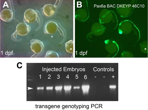
EGFP expression typically seen in zebrafish embryos transiently transgenic for the dualreporter BAC at 1 dpf. (A) These eight embryos were randomly chosen from a group of 80 embryos. (B) In this group, seven embryos exhibited comparable EGFP fluorescence, and one had only a few EGFP-expressing cells in the eye (*). In this group, 74 embryos (>90%) exhibited EGFP expression. None of these embryos exhibited EGFP expression in cells outside of the normal domain of Pax6a expression. (C) PCR genotyping analysis indicates that >90% (n=94/100 embryos; three independent trials) of the injected embryos retain the BAC transgene after 1 day of development. Eight representative examples are shown: six are from embryos injected with the BAC transgene (1-6), and two are from uninjected controls (-). +, positive control. |

The zebrafish Pax6a BAC transgene exhibits differential promoter activity in the adult mouse retina. DsRed is expressed by a subset of EGFP+ cells in the adult mouse retina. Both EGFP and DsRed were visualized by indirect immunofluorescence (EGFP, green; DsRed, red). Arrowheads denote examples of EGFP+/DsRed- cells. Horizontal and ganglion cells express EGFP but not DsRed. DsRed is expressed by a subset of EGFP+ cells in the both the GCL and INL. Marker analyses presented in this study showed that GABAergic amacrine cells express both DsRed and EGFP; DsRed expressing neurites tend to localize to the middle strata of the IPL (arrow in B). A subset of bipolar cells also coexpresses DsRed and EGFP. |
Reprinted from Developmental Biology, 307(2), Lakowski, J., Majumder, A., and Lauderdale, J.D., Mechanisms controlling Pax6 isoform expression in the retina have been conserved between teleosts and mammals, 498-520, Copyright (2007) with permission from Elsevier. Full text @ Dev. Biol.