
Fig. 7
- ID
- ZDB-FIG-070810-22
- Publication
- Lakowski et al., 2007 - Mechanisms controlling Pax6 isoform expression in the retina have been conserved between teleosts and mammals
- Other Figures
- All Figure Page
- Back to All Figure Page
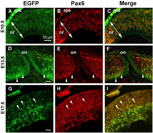
Zebrafish Pax6a P0/P1 promoters are active in Pax6-expressing cells in the developing mouse retina. Sections cut through the eyes of transgenic mouse embryos at E10.5 (A–C), E13.5 (D–F), or E17.5 (G–I). Both EGFP (A, D, G) and endogenous Pax6 (B, E, H) were visualized by indirect immunofluorescence (EGFP, green; Pax6, red). (C, F, I) Merged images. EGFP and Pax6 are coexpressed in cells in the neural retina (nr). At E10.5, EGFP expression replicated mouse Pax6. At both E13.5 (D–F) and E17.5 (G–I), EGFP was more strongly expressed in putative differentiating neurons (arrowheads) than in neuroepithelial cells. At E13.5 EGFP expression in retinal ganglion cell axons is visible within the optic nerve (on). Arrowheads in D–F denote ganglion cells with axonal projections into the developing optic nerve. Scale bars = 25 μm; bar in panel A applies to panels B–C; bar in panel D applies to panels E–I. |
Reprinted from Developmental Biology, 307(2), Lakowski, J., Majumder, A., and Lauderdale, J.D., Mechanisms controlling Pax6 isoform expression in the retina have been conserved between teleosts and mammals, 498-520, Copyright (2007) with permission from Elsevier. Full text @ Dev. Biol.