
|
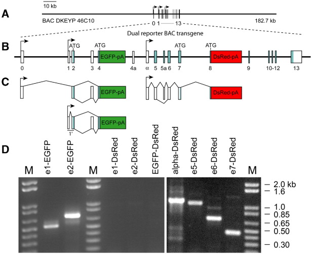
Fig. 2 Structure and transcript analysis of the zebrafish Pax6a dual-reporter BAC transgene. (A) Schematic representation of zebrafish BAC DKEY-46C10 showing the location of the Pax6a transcript unit within the BAC. Black boxes denote exons, and bent arrows denote promoters P0, P1, and Palpha. (B) The zebrafish Pax6a BAC was modified by insertion of an enhanced green fluorescent protein reporter cassette (EGFP pA) in-frame with the evolutionarily conserved ATG in exon 4. A Discosoma red fluorescent protein reporter cassette (DsRed-pA) was inserted in-frame into the conserved ATG in exon 8. (C) Whereas transcripts that initiate from the P0 and P1 promoters encode for EGFP, Palpha-initiated transcripts encode for DsRed. (D) Analysis of transcripts from the BAC transgene. RNA was prepared from 3-day old embryos transiently transgenic for the dual-reporter BAC and analyzed by RT-PCR. Transcripts with exon 1 (e1) or exon 2 (e2) also included EGFP, but not DsRed. Transcripts with exons alpha, 5 (e5), 6 (e6), or 7 (e7) also included DsRed. No P1- or P0-initiated transcripts contained DsRed.
Reprinted from Developmental Biology, 307(2), Lakowski, J., Majumder, A., and Lauderdale, J.D., Mechanisms controlling Pax6 isoform expression in the retina have been conserved between teleosts and mammals, 498-520, Copyright (2007) with permission from Elsevier. Full text @ Dev. Biol.