
- Title
-
Role of Fyn kinase in signaling associated with epiboly during zebrafish development
- Authors
- Sharma, D., Holets, L., Zhang, X., and Kinsey, W.H.
- Source
- Full text @ Dev. Biol.
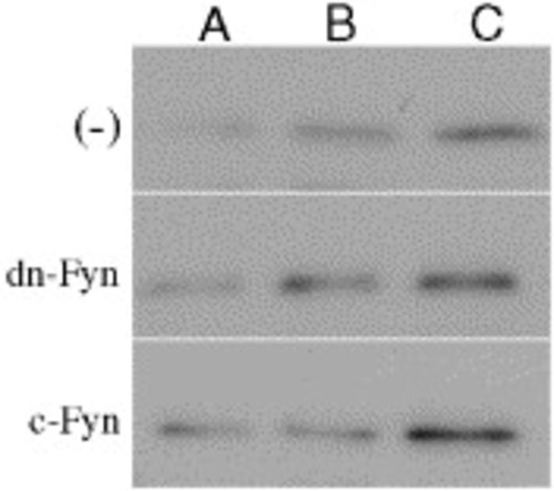
Expression of Fyn kinase following mRNA injection. The level of Fyn protein expression was monitored by Western blot analysis using an anti-peptide antibody specific for Fyn kinase (Fyn-3 antibody from Santa Cruz Biotechnology, Santa Cruz, CA). Membrane fractions were prepared and Western blots were performed from groups of 20 embryos as previously described (Wu and Kinsey, 2003). Samples containing identical amounts of protein from control, uninjected embryos (-), from embryos injected with 230 pg of FynK299M (dn-Fyn), or c-Fyn mRNA (c-Fyn) were collected at 3.3 hpf (high stage, A), 3.8 hpf (sphere stage, B), or at 4.5 hpf when controls were at 25% epiboly (C). EXPRESSION / LABELING:
|
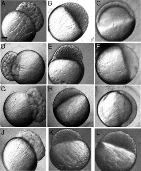
Morphology of embryos arrested following FynK299M mRNA or GST-Fyn-SH2 injection. Zygotes were injected with 230–250 pg of mRNA encoding c-Fyn (A–C) or FynK299M (D–F), or a mixture of c-Fyn (230 pg) and of FynK299M (270 pg), then incubated in embryo medium at 28°C. Zygotes injected with the GST-Fyn-SH2 fusion protein (1.3 pmol) also ceased development at the onset of epiboly (J, K, L). They were examined at 2.5 hpf (A, D, G, J), 4.0 hpf (B, E, H, K), and at 5.25 hpf (C, F, I, L). Magnification is indicated by the bar in panel A which indicates 100 μm. |
Morphology and distribution of cell nuclei in normal and blocked embryos. Embryos were injected with c-Fyn (A–D) or FynK299M mRNA (E–H) as in Fig. 4. When the control embryos reached 50% epiboly, the embryos were fixed in 2% glutaraldehyde, permeabilized with 0.1% TX 100, and stained with DAPI. The embryos were then examined with bright field (A, C, E, G), or with fluorescence microscopy to localize the DAPI-stained nuclei (B, D, F, H). Arrows indicate the position of YSL nuclei. Magnification is indicated by the bar which indicates 100 μm. |
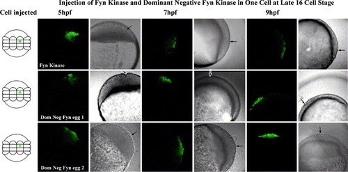
Effect of FynK299M on the ability of blastomeres to participate in epiboly. Embryos were collected at the 16-cell stage and single blastomeres were injected with a mixture of calcium green-dextran and mRNA (4.5 pg). The embryos were cultured at 28°C and examined by confocal fluorescence microscopy at 5, 7, and 9 hpf. The embryo shown in the upper panels was injected with c-Fyn mRNA and the fluorescent cells migrated toward the vegetal pole with the advancing blastoderm. The embryos shown in the middle and lower panels were injected with FynK299M mRNA and in these cases, the fluorescent cells remained near the animal pole. |
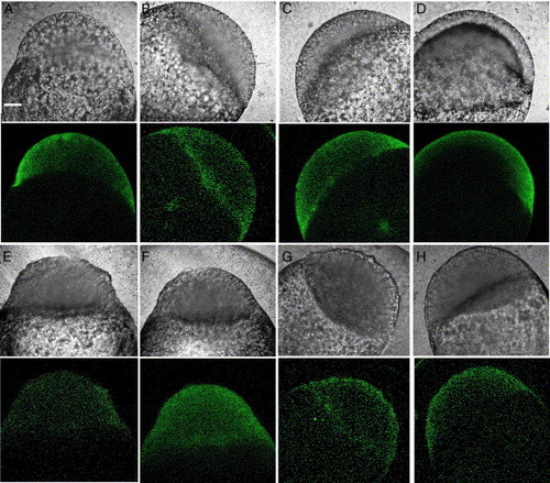
Morphology of calcium green fluorescence in normal and FynK299M-injected embryos. Embryos injected with calcium green-dextran and either c-Fyn mRNA (A–D) or FynK299M mRNA (E–H) as in Fig. 8 were imaged using a transmission detector to produce a ‘bright field’ image (upper panels) and separate images were recorded with the green filter (lower panels) to demonstrate calcium green fluorescence. Images were taken when the control embryo was at the high stage (3.3 hpf, A and E), sphere stage (3.8 hpf, B and F), dome stage (4.55 hpf, C and G), and at 50% epiboly (5.5 hpf, D and H). Magnification is indicated by the white bar which represents 100 μm. (For interpretation of the references to colour in this figure legend, the reader is referred to the web version of this article.) |
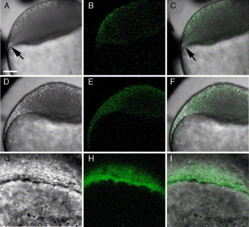
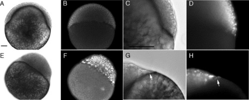
Morphology of the marginal region of the blastodisc in normal zebrafish embryos. Embryos developed from zygotes injected with calcium green-dextran were imaged by confocal microscopy to obtain ‘bright field’ and green fluorescence images demonstrating the distribution of calcium green fluorescence in the embryo. An embryo was imaged at sphere stage (A–C) and at about 20% epiboly (D–I). Images A–F are focused through the equator of the embryo, while in images G–I, the focal plane passed tangentially through the leading edge of the advancing blastoderm. Arrows in panels A and C indicate the position of the YSL. Magnification is indicated by the bar which represents 100 μm. |

Effect of FynK299M mRNA and GST-Fyn-SH2 protein on the localization of boz, goosecoid, mezzo, and bon/mixer. Embryos resulting from zygotes injected with c-Fyn mRNA (230 pg) as a control (A, D, G, J) or with 230 pg of FynK299M mRNA (B, E, H, K) or with GST-Fyn-SH2 (2 pmol) (C, F, I, L) were incubated in embryo medium at 28°C, then fixed at 5 hpf and processed for in situ hybridization as described in Materials and methods. Magnification is indicated by the bar which represents 100 μm. |
Reprinted from Developmental Biology, 285(2), Sharma, D., Holets, L., Zhang, X., and Kinsey, W.H., Role of Fyn kinase in signaling associated with epiboly during zebrafish development, 462-476, Copyright (2005) with permission from Elsevier. Full text @ Dev. Biol.