
FIGURE
Fig. 5
- ID
- ZDB-FIG-070920-45
- Publication
- Sharma et al., 2005 - Role of Fyn kinase in signaling associated with epiboly during zebrafish development
- Other Figures
- All Figure Page
- Back to All Figure Page
Fig. 5
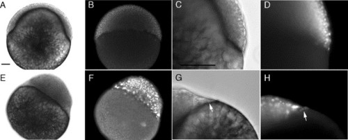
Morphology and distribution of cell nuclei in normal and blocked embryos. Embryos were injected with c-Fyn (A–D) or FynK299M mRNA (E–H) as in Fig. 4. When the control embryos reached 50% epiboly, the embryos were fixed in 2% glutaraldehyde, permeabilized with 0.1% TX 100, and stained with DAPI. The embryos were then examined with bright field (A, C, E, G), or with fluorescence microscopy to localize the DAPI-stained nuclei (B, D, F, H). Arrows indicate the position of YSL nuclei. Magnification is indicated by the bar which indicates 100 μm. |
Expression Data
Expression Detail
Antibody Labeling
Phenotype Data
Phenotype Detail
Acknowledgments
This image is the copyrighted work of the attributed author or publisher, and
ZFIN has permission only to display this image to its users.
Additional permissions should be obtained from the applicable author or publisher of the image.
Reprinted from Developmental Biology, 285(2), Sharma, D., Holets, L., Zhang, X., and Kinsey, W.H., Role of Fyn kinase in signaling associated with epiboly during zebrafish development, 462-476, Copyright (2005) with permission from Elsevier. Full text @ Dev. Biol.