
- Title
-
Genetic locus half baked is necessary for morphogenesis of the ectoderm
- Authors
- McFarland, K.N., Warga, R.M., and Kane, D.A.
- Source
- Full text @ Dev. Dyn.
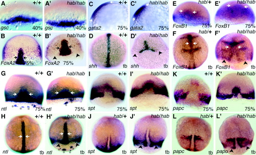
Specification in hab mutants before and during gastrulation. In the following experiments, all hab-/+ embryos are zygotic maternal dominant (ZMD) embryos; during these stages, in situ analysis cannot distinguish between the wild-type and dominant phenotypes. Embryos were genotyped after completion of photography as outlined in the Experimental Procedures section. All embryos are dorsal views with the animal pole to the top, and arrows indicate the width of the axial expression pattern, except C, which is a side view with dorsal to the right and arrows indicate the anteroposterior extent of expression. Arrowheads indicate the edge of the blastoderm margin in mutant embryos. A: Expression of goosecoid, 40% epiboly, in wild-type and hab-/-, dorsal view. B: Expression of FoxA2/axial/forkhead 1, 75% epiboly in wild-type and hab-/-. C: Expression of gata2, 75% epiboly in wild-type and hab-/- (C). Arrows indicate the expression domain. D: sonic hedgehog, tail bud stage in wild-type and hab-/-. Note the absence of expression in the anterior neural plate of the mutant. E,F: FoxB1/mariposa/forkhead 3, 75% epiboly and tail bud stage in ZMD hab-/+ and hab-/-. E′ and F′: Arrows indicate widening of the midline in the mutants. F′: Note gaps in the midline and absence of anterior forebrain expression. G,H: no tail, 75% epiboly and tail bud stage in wild-type and hab-/-. Ectopic forerunner cell clusters are indicated by asterisks. Wild-type embryos in G are from hab-/+ mothers. I,J: spadetail, 75% epiboly and tail bud stage in wild-type, ZMD hab-/+ and hab-/-. K,L: paraxial protocadherin, 75% epiboly and tail bud stage in wild-type, ZMD hab-/+and hab-/-. Note that, in the hab-/- mutants, expression of both spadetail and paraxial protocadherin in the prechordal plate is retained longer. EXPRESSION / LABELING:
|
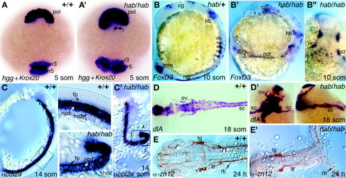
Differentiation in hab mutants after gastrulation. Embryos were genotyped after completion of photography as outlined in the Experimental Procedures section. A: Expression of hatching gland gene and Krox20, five-somite stage in wild-type (A) and hab-/-(A′), dorsoanterior view. Note ectopic hatching gland cell (arrow) below pollster in the mutant. B: FoxD3/forkhead 6, 10-somite stage in zygotic maternal dominant hab-/+ and hab-/-. Side view except for B′, which is a dorsal view. Note weak expression in mutant tail bud. Dorsal view shows bilateral somites (indicated by asterisks) forming on either side of the germ ring. C: alpha-collagen2a, 14-somite stage in wild-type (side view) and hab-/- (dorsal view of kinked notochord). C′: Higher magnification image of the boxed regions. D: delta A,18-somite stage in wild-type (dorsal view, D) and hab-/- (dorsal view and side view, D′). E: Zn12 antibody, 24 hr in wild-type (E) and rare hab-/- escaper (E′), dorsal view. Note bifurcation of axis in mutant. cg, cranial ganglia; fp, floor plate; hb, hindbrain; hcd, hypochord; llf, lateral longitudinal fascicle; mb, midbrain; ncd, notochord; ncr, neural crest; os, optic stalk; ov, otic vesicles; pol, polster; r3, rhombomere3; r5, rhombomere5; rb, Rohan-Beard cells; sc, spinal cord; tbm, tail bud mesenchyme; tg, trigeminal. Arrows indicate the width of the axis. |