
- Title
-
Growth cones utilize both widespread and local directional cues in the zebrafish brain
- Authors
- Kanki, J.P. and Kuwada, J.Y.
- Source
- Full text @ Dev. Biol.
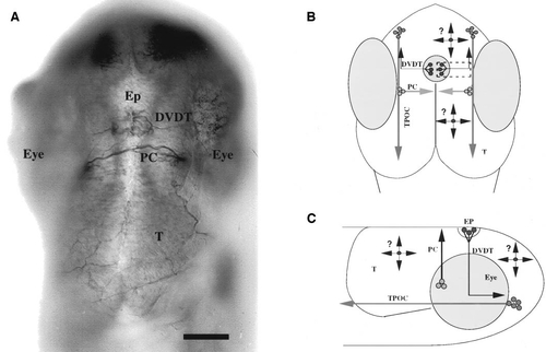
Epiphysial neurons are located in an accessible site. (A) Dorsal view of the head of a 28 hpf embryo showing that the epiphysial neurons are located at the base of the epiphysis. Anterior is up. Axons were labeled with an antibody against acetylated α-tubulin. The epiphysial axons on each side of the epiphysis (Ep), which is located at the dorsal midline, run laterally and ventrally to form the dorsoventral diencephalic tract (DVDT). Posterior to the DVDT is the posterior commissure (PC) that runs just anterior to the tectum (T). Scale is 50 μm. (B) Schematic diagram illustrating the rationale of the transplantation experiments on a dorsal view of the embryo. Axons of epiphysial neurons extend from the epiphysis (large circle at the midline) to pioneer the DVDT that extends along the surface of the diencephalon. The axons then turn anteriorly into the tract of the postoptic commissure (TPOC). The TPOC is pioneered by neurons (anterior cluster of circles) in the anterior and ventral diencephalon. The PC is pioneered by dorsally directed axons from neurons located in the anterior tegmentum (cluster of circles just medial to the eyes). The black circles with arrows and question marks represent ectopically transplanted epiphysial neurons and some of the possible directions that their axons could extend in the brain. (C) Schematic diagram illustrating the rationale of the transplantation experiments on a lateral view of the embryo. Anterior is to the right and dorsal is up. |
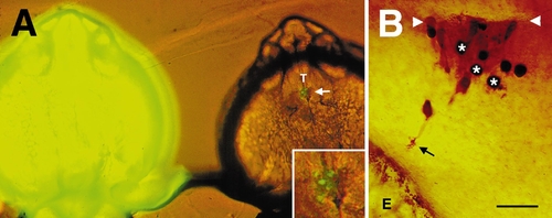
Epiphysial neurons extend axons ventrally when transplanted to ectopic sites in the brain. (A) Micrograph showing a live, FITC–dextran-labeled donor (left) and a live, unlabeled host (right) embryo at 24 hpf in which epiphysial neurons (arrow) had been isochronically transplanted at 20 hpf. Inset shows an enlargement of the labeled transplanted cells. Anterior is up; T marks the tectum. (B) A dorsal view of the tectum in another 24 hpf embryo showing that epiphysial neurons project growth cones (arrow) that extend ventrally when transplanted into the tectum 4 h earlier. Here all donor cells are marked brown by labeling them with an antibody against FITC–dextran. Many of the transplanted cells (some marked with an asterisk) that do not extend axons are presumably nonneuronal cells that were transplanted along with the epiphysial cells. Arrowheads denote the dorsal midline of the tectum and E the eye. Scale is 150 μm in A and 30 μm in B. |

Epiphysial axons extend ventrally even in the vicinity of the posterior commissure (PC) that contains many dorsally extending axons. (A) Dorsal view of the DVDT and PC axons labeled with anti-tubulin and visualized using an alkaline phosphatase reaction. Anterior is up; Ep denotes the epiphysis; scale, 25 μm. (B) Lateral view of the host PC (arrowheads) labeled blue as in A and a donor epiphysial axon (arrow) labeled with anti-FITC and marked by the brown peroxidase reaction product. The donor axon is extending ventrally along the host PC. Anterior is to the left and dorsal is up; scale, 25 μm. (C) Schematic diagram summarizing the pathways taken by the axons of the donor epiphysial neurons when transplanted onto or near the PC. Neurons with ventrally extending axons are designated with filled circles and those with dorsally extending axons are designated with open circles. The latter are the same neurons shown as open circles in Fig. 3B. The donor neuron on the left side first extended dorsally and then turned and extended ventrally. |
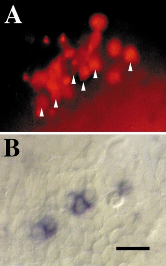
Epiphysial neurons which normally express islet-1 retain expression of islet-1 following transplantation to ectopic sites. (A) Lateral view of the embryo showing the TRITC–dextran-labeled donor cells in the host tectum. Arrowheads mark donor cells that expressed islet-1 mRNA that can be seen in B. (B) Same view as in A showing that some of the donor cells express islet-1 as determined by in situ hybridization to an islet-1 riboprobe. These cells are designated by arrowheads in A. Anterior is to the left and dorsal is up; scale, 25 μm. |
Reprinted from Developmental Biology, 219(2), Kanki, J.P. and Kuwada, J.Y., Growth cones utilize both widespread and local directional cues in the zebrafish brain, 364-372, Copyright (2000) with permission from Elsevier. Full text @ Dev. Biol.