
|
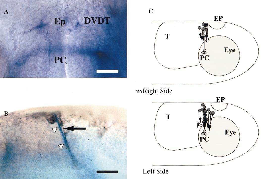
Fig. 4 Epiphysial axons extend ventrally even in the vicinity of the posterior commissure (PC) that contains many dorsally extending axons. (A) Dorsal view of the DVDT and PC axons labeled with anti-tubulin and visualized using an alkaline phosphatase reaction. Anterior is up; Ep denotes the epiphysis; scale, 25 μm. (B) Lateral view of the host PC (arrowheads) labeled blue as in A and a donor epiphysial axon (arrow) labeled with anti-FITC and marked by the brown peroxidase reaction product. The donor axon is extending ventrally along the host PC. Anterior is to the left and dorsal is up; scale, 25 μm. (C) Schematic diagram summarizing the pathways taken by the axons of the donor epiphysial neurons when transplanted onto or near the PC. Neurons with ventrally extending axons are designated with filled circles and those with dorsally extending axons are designated with open circles. The latter are the same neurons shown as open circles in Fig. 3B. The donor neuron on the left side first extended dorsally and then turned and extended ventrally.
Reprinted from Developmental Biology, 219(2), Kanki, J.P. and Kuwada, J.Y., Growth cones utilize both widespread and local directional cues in the zebrafish brain, 364-372, Copyright (2000) with permission from Elsevier. Full text @ Dev. Biol.