
FIGURE
Fig. 6
- ID
- ZDB-FIG-080522-24
- Publication
- Kanki et al., 2000 - Growth cones utilize both widespread and local directional cues in the zebrafish brain
- Other Figures
- All Figure Page
- Back to All Figure Page
Fig. 6
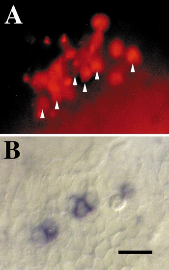
Epiphysial neurons which normally express islet-1 retain expression of islet-1 following transplantation to ectopic sites. (A) Lateral view of the embryo showing the TRITC–dextran-labeled donor cells in the host tectum. Arrowheads mark donor cells that expressed islet-1 mRNA that can be seen in B. (B) Same view as in A showing that some of the donor cells express islet-1 as determined by in situ hybridization to an islet-1 riboprobe. These cells are designated by arrowheads in A. Anterior is to the left and dorsal is up; scale, 25 μm. |
Expression Data
Expression Detail
Antibody Labeling
Phenotype Data
Phenotype Detail
Acknowledgments
This image is the copyrighted work of the attributed author or publisher, and
ZFIN has permission only to display this image to its users.
Additional permissions should be obtained from the applicable author or publisher of the image.
Reprinted from Developmental Biology, 219(2), Kanki, J.P. and Kuwada, J.Y., Growth cones utilize both widespread and local directional cues in the zebrafish brain, 364-372, Copyright (2000) with permission from Elsevier. Full text @ Dev. Biol.