
- Title
-
The expression pattern of the mouse receptor tyrosine kinase gene MDK1 is conserved through evolution and requires Hoxa-2 for rhombomere-specific expression in mouse embryos
- Authors
- Taneja, R., Thisse, B., Rijli, F.M., Thisse, C., Bouillet, P., Dollé, P., and Chambon, P.
- Source
- Full text @ Dev. Biol.
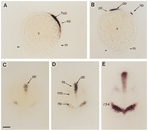
Early expression pattern of ZDK1 during gastrulation. (A) At 60% epiboly, expression is seen in the hypoblast but not in the epiblast. (B) At 80% epiboly, additional expression is detected in the presumptive hindbrain region. (C) Dorsal view of embryo shown in B. (D and E) Dorsal views of embryos at 100% epiboly (D) and bud stage (E) showing ZDK1 expression in hindbrain, in the forebrain– midbrain boundary, and in the prechordal plate mesendoderm which underlies the ventral forebrain. Abbreviations: ep, epiblast; fb, forebrain; hb, hindbrain; hyp, hypoblast; m, margin; mb, midbrain; pil, pillow; pp, prechordal plate; r3-4, rhombomeres 3 and 4; y, yolk. (A and B) Side view, dorsal is to the right, anterior is to the top. (C–E) Dorsal view, anterior is up. Scale bar: A–D, 25 μm; E, 50 μm. |
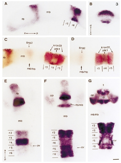
Expression of ZDK1 in the hindbrain and the forebrain. (A) Lateral view of an 8 somite embryo. (B) Thick dorsoventral section at the level of r4 of the same embryo shown in A. (C) Lateral view and (D) dorsal view of an 8 somite embryo, double labeled with Krox- 20 (in red), Engrailed 2 (Eng 2, in red), and ZDK1 (in brown–black). (E–G) Dorsal views of 10 somite (E) 20 somite (F), and 24 hr (G) embryos. Abbreviations: a, anterior; d, dorsal; di, diencephalon; fb, forebrain; fb/mb, forebrain–midbrain junction; mb, midbrain; mb/hb, midbrain– hindbrain junction; op, optic vesicle; ov, otic vesicle; p, posterior; r2– r6, rhombomeres 2–6; t, telencephalon; v, ventral. Scale bar: A– F, 50 μm; G, 25 μm. EXPRESSION / LABELING:
|
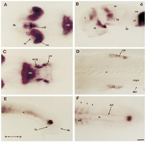
ZDK1 expression at later stages of development. (A) 36 hr embryo viewed dorsally showing strong expression in the posterior retina, lens, and ventral forebrain. (B) Lateral view of the same embryo as in A after removing the eye. (C) Posterior domains of expression of the same embryo shown in A. Optical cross section at the level of accoustic ganglia and otic vesicle. (D) Optical, horizontal cross section of a 36 hr embryo at the level of the caudal lateral line primodium. (E and F) Caudal expression in the lateral line primordium at the tip of the notochord of a 14 somite embryo. (E) Lateral view; (F) dorsal view. Abbreviations: a, anterior; acg, accoustic ganglia; ad, adaxial cells; ce, cerebellum; der, dermis; di, diencephalon; fb, ventral forebrain; fp, floor plate; hb, hindbrain; Kv, Kuppfer vesicle; le, lens; llp, lateral line primordium; myo, myotome; n, notochord; ov, otic vesicle; p, posterior; r, rhombomere; re, retina; so, somite; t, telencephalon; te, tectum; tb, tailbud; tg, tegmentum; vdi, ventral diencephalon. Arrowheads indicate the positions of the somitic furrows. Scale bar: A–C, 50 μm; D, 100 μm. Anterior is to the left. EXPRESSION / LABELING:
|
Reprinted from Developmental Biology, 177(2), Taneja, R., Thisse, B., Rijli, F.M., Thisse, C., Bouillet, P., Dollé, P., and Chambon, P., The expression pattern of the mouse receptor tyrosine kinase gene MDK1 is conserved through evolution and requires Hoxa-2 for rhombomere-specific expression in mouse embryos, 397-412, Copyright (1996) with permission from Elsevier. Full text @ Dev. Biol.