
|
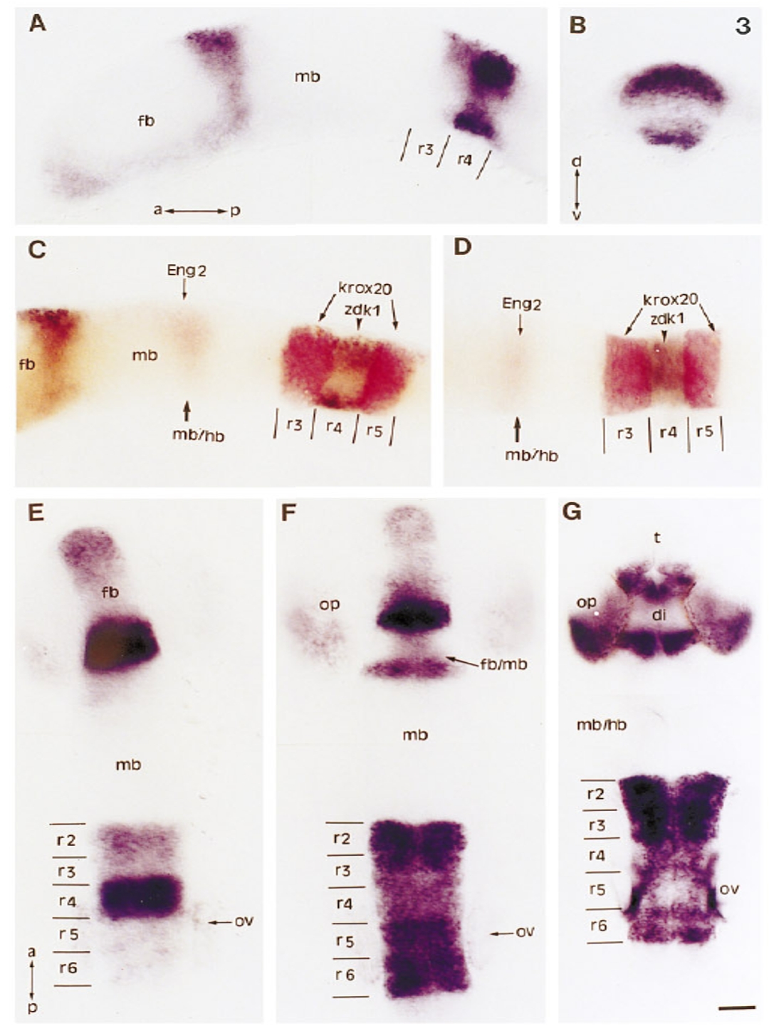
Fig. 3 Expression of ZDK1 in the hindbrain and the forebrain. (A) Lateral view of an 8 somite embryo. (B) Thick dorsoventral section at the level of r4 of the same embryo shown in A. (C) Lateral view and (D) dorsal view of an 8 somite embryo, double labeled with Krox- 20 (in red), Engrailed 2 (Eng 2, in red), and ZDK1 (in brown–black). (E–G) Dorsal views of 10 somite (E) 20 somite (F), and 24 hr (G) embryos. Abbreviations: a, anterior; d, dorsal; di, diencephalon; fb, forebrain; fb/mb, forebrain–midbrain junction; mb, midbrain; mb/hb, midbrain– hindbrain junction; op, optic vesicle; ov, otic vesicle; p, posterior; r2– r6, rhombomeres 2–6; t, telencephalon; v, ventral. Scale bar: A– F, 50 μm; G, 25 μm.
Reprinted from Developmental Biology, 177(2), Taneja, R., Thisse, B., Rijli, F.M., Thisse, C., Bouillet, P., Dollé, P., and Chambon, P., The expression pattern of the mouse receptor tyrosine kinase gene MDK1 is conserved through evolution and requires Hoxa-2 for rhombomere-specific expression in mouse embryos, 397-412, Copyright (1996) with permission from Elsevier. Full text @ Dev. Biol.