
|
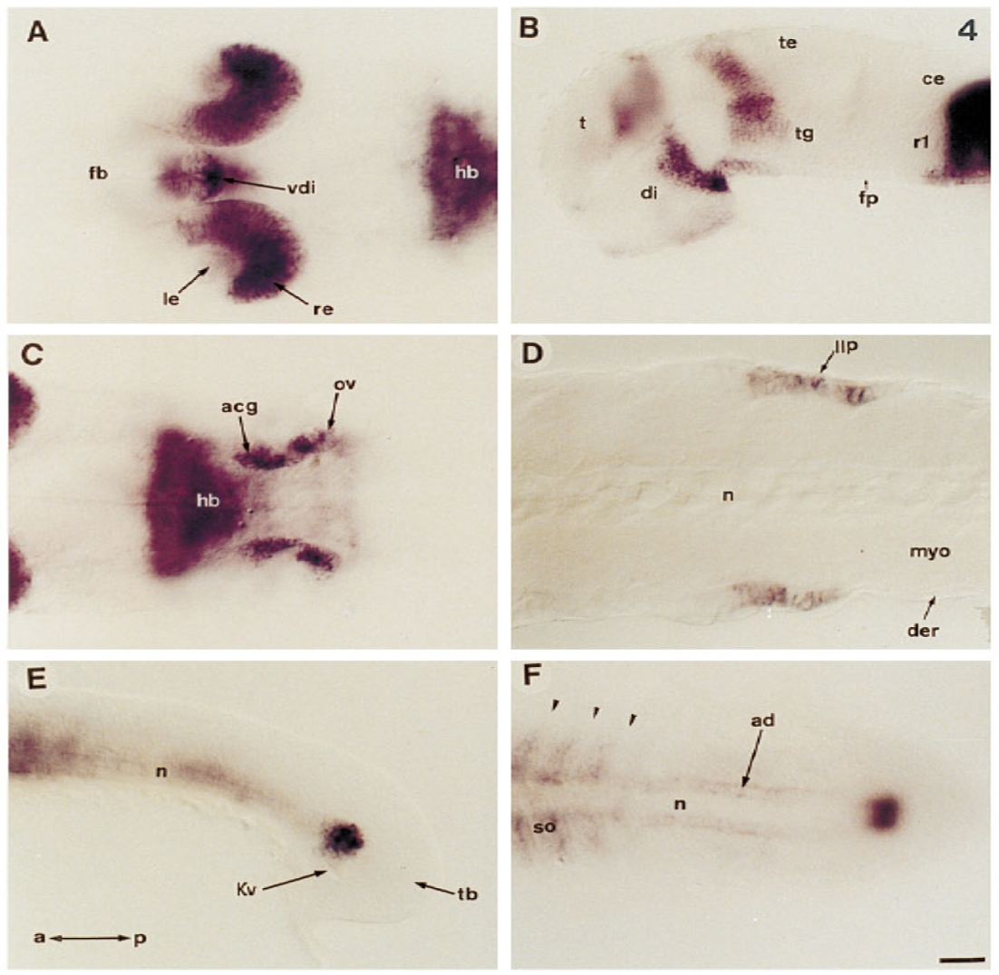
Fig. 4 ZDK1 expression at later stages of development. (A) 36 hr embryo viewed dorsally showing strong expression in the posterior retina, lens, and ventral forebrain. (B) Lateral view of the same embryo as in A after removing the eye. (C) Posterior domains of expression of the same embryo shown in A. Optical cross section at the level of accoustic ganglia and otic vesicle. (D) Optical, horizontal cross section of a 36 hr embryo at the level of the caudal lateral line primodium. (E and F) Caudal expression in the lateral line primordium at the tip of the notochord of a 14 somite embryo. (E) Lateral view; (F) dorsal view. Abbreviations: a, anterior; acg, accoustic ganglia; ad, adaxial cells; ce, cerebellum; der, dermis; di, diencephalon; fb, ventral forebrain; fp, floor plate; hb, hindbrain; Kv, Kuppfer vesicle; le, lens; llp, lateral line primordium; myo, myotome; n, notochord; ov, otic vesicle; p, posterior; r, rhombomere; re, retina; so, somite; t, telencephalon; te, tectum; tb, tailbud; tg, tegmentum; vdi, ventral diencephalon. Arrowheads indicate the positions of the somitic furrows. Scale bar: A–C, 50 μm; D, 100 μm. Anterior is to the left.
Reprinted from Developmental Biology, 177(2), Taneja, R., Thisse, B., Rijli, F.M., Thisse, C., Bouillet, P., Dollé, P., and Chambon, P., The expression pattern of the mouse receptor tyrosine kinase gene MDK1 is conserved through evolution and requires Hoxa-2 for rhombomere-specific expression in mouse embryos, 397-412, Copyright (1996) with permission from Elsevier. Full text @ Dev. Biol.