
- Title
-
Mutations affecting cell fates and cellular rearrangements during gastrulation in zebrafish
- Authors
- Solnica-Krezel, L., Stemple, D.L., Mountcastle-Shah, E., Rangini, Z., Neuhauss, S.C., Malicki, J., Schier, A.F., Stainier, D.Y., Zwartkruis, F., Abdelilah, S., and Driever, W.
- Source
- Full text @ Development
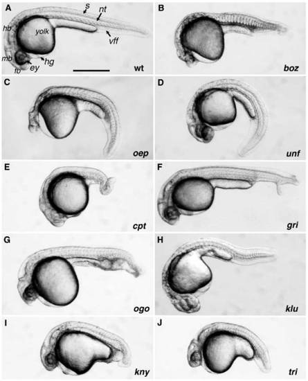
The phenotypes of gastrulation mutants at day one of development. Dissecting microscope images of live embryos: (A) Wild type (wt); (B) bozozokm168, (C) one-eyedpinheadm134, (D) uncle freddym768, (E) captain hookm52, (F) grinchm100, (G) ogonm60, (H) kluskam472, (I) knypekm119 and (J) trilobitem209 mutant embryos. ey, eye; fb, forebrain; hb, hindbrain; hg, hatching gland; mb, midbrain; nt, notochord; vff, ventral fin fold. Scale bar, 0.5 mm. PHENOTYPE:
|
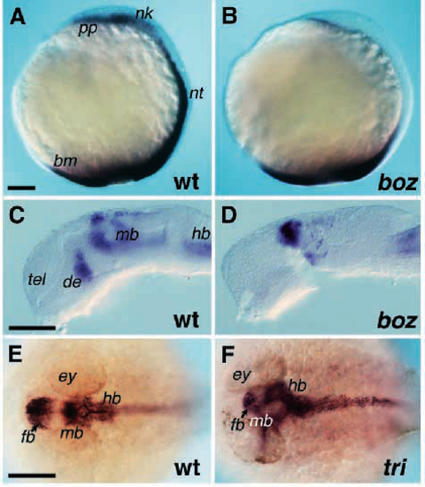
Expression patterns of cell type and region-specific genes during development of mutants affecting dorsal fates. (A,B) Expression of gsc and ntl RNA in wild-type (wt) (A) and bozm168 (B) mutant embryos at the bud stage (9.5 hpf). pp, gsc expression domain in prechordal plate; nk, gsc expression domain in neural keel; nt, ntl expression domain in chordamesoderm; bm, ntl expression domain in the blastoderm margin. (C,D) Expression pattern of hlx1 RNA in wild-type (C) and bozm168 mutant embryos at 30 hpf; de, mb, expression domain in diencephalon and midbrain, respectively. (E,F) Expression of shh RNA in wild-type (E) and trim209 mutant embryo at 30 hpf. ey, eye; fb, forebrain; hb, hindbrain; mb, midbrain. Scale bar, 0.1 mm. EXPRESSION / LABELING:
PHENOTYPE:
|

ZFIN is incorporating published figure images and captions as part of an ongoing project. Figures from some publications have not yet been curated, or are not available for display because of copyright restrictions. |
|
ZFIN is incorporating published figure images and captions as part of an ongoing project. Figures from some publications have not yet been curated, or are not available for display because of copyright restrictions. PHENOTYPE:
|
|
ZFIN is incorporating published figure images and captions as part of an ongoing project. Figures from some publications have not yet been curated, or are not available for display because of copyright restrictions. PHENOTYPE:
|
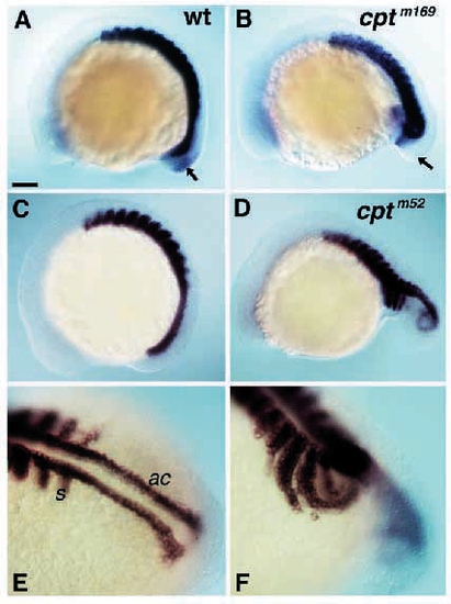
Decrease in the expression of ventroposterior marker eve1, and lateral expansion of a somitic marker myoD in cpt mutant embryos during somitogenesis. (A,C,E) Wild type (wt); (B) cptm169 and (D,F) cptm52 mutant embryos. (A,B) Expression of myoD and eve1, eve1 expression domain in the tail is indicated by an arrow. (C-F) Expression of myoD mRNA. (A-D) Lateral views, anterior towards the left and dorsal towards the top; (E,F) Dorsolateral view of developing tail. s, somites; ac, adaxial cells. Scale bar, 0.1 mm. EXPRESSION / LABELING:
|
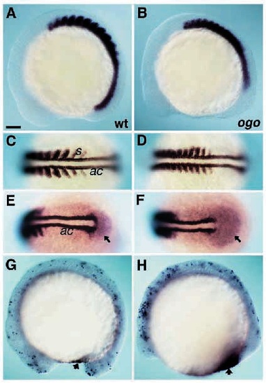
Expression patterns of cell type and region-specific genes at the 10 somite stage in ogom60 mutants affected in formation of ventral and posterior fates. (A,C,E) Wild type (wt); (B,D,F) ogom60 mutant emryo. (A-D) In situ hybridization of myoD mRNA; (E,F) Expression of myoD and eve1 mRNA (arrow). (G,H) Visualization of dying cells in ogom60 mutants at the 13 somite stage. A ventral region with increased numbers of dying cells is indicated by an arrow. (G,H) Detection of apoptotic cells in wild-type (G) and ogo (H) mutant embryos at the 13 somite stage. Arrow indicates a region with increased number of dying cells. (A,B) Lateral views, anterior towards the left and dorsal towards the top; (C,D) Dorsal view; (E,F) Dorsoposterior view. s, somites, ac, adaxial cells. Scale bar, 0.1 mm. EXPRESSION / LABELING:
PHENOTYPE:
|
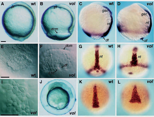
Changes in cellular rearrangements and patterns of gene expression during development of the volm712 mutants. (A,C,E,G,K) Wild type (wt); (B,D,F,H,I,J,L) volm712 mutant embryo. (A,B) Dissecting microscope images at the tail bud stage. Lateral view, dorsal toward the right. (E,F,I) Nomarski images at the tail bud stage, 30 minutes after the yolk plug closure in wild-type embryos. pl, polster; dcm, margin of deep cells; df, dorsal forerunner cells. (J) Dissecting microscope image of a volm712 mutant embryo at 10 hours. Embryo is in chorion, blastoderm is disintegrating. (C,D,G,H) Expression of gsc and ntl mRNA at 90% epiboly. (K,L) Expression of axial mRNA at 90% epiboly. nt, notochord expression domain of ntl; df, dorsal forerunner expression domain of ntl. Scale bars, 0.1 mm. EXPRESSION / LABELING:
PHENOTYPE:
|
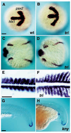
Changes in expression patterns of cell type and region-specific genes in mutants affected in convergence and extension. (A,C,E,G) Wild type (wt); (B,D,F) trim209 and (H) knym119 mutant embryos. (A,B) Expression of hlx1 and pax2 mRNA at the tail bud stage, view of developing head region. Anterior is towards the left. (C,D) Expression of pax2 at the tail bud stage. Dorsal view. (E,F) Expression of myoD mRNA at the 10 somite stage. Dorsal view, anterior toward the left. (G,H) Expression of eve1 mRNA in tail at 1 dpf. Note ectopic expression in the mutant embryo. Scale bar, 0.1 mm. |
|
ZFIN is incorporating published figure images and captions as part of an ongoing project. Figures from some publications have not yet been curated, or are not available for display because of copyright restrictions. PHENOTYPE:
|