
- Title
-
Mutations affecting neural survival in the zebrafish Danio rerio
- Authors
- Abdelilah, S., Mountcastle-Shah, E., Harvey, M., Solnica-Krezel, L., Schier, A.F., Stemple, D.L., Malicki, J., Neuhauss, S.C., Zwartkruis, F., Stainier, D.Y., Rangini, Z., and Driever, W.
- Source
- Full text @ Development

ZFIN is incorporating published figure images and captions as part of an ongoing project. Figures from some publications have not yet been curated, or are not available for display because of copyright restrictions. PHENOTYPE:
|
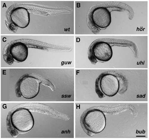
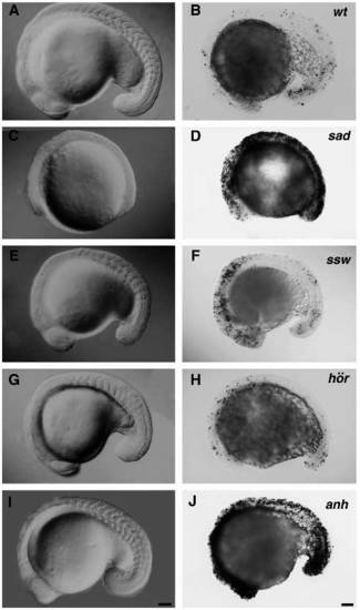
Mutants of the spacehead and miscellaneous early degeneration classes at 28 hpf. (A) Wild-type. (B) hörnlem274. (C) gumowym585. (D) uchu hikoushim172. (E) sideswipem762. (F) stop and dropm367. (G) anhalterm767. (H) bubblesm450. Lateral view of live embryos. Transmitted light, anterior to the left. Scale bar, 250 µm. |
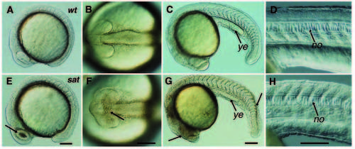
Early development of spacehead mutant saturnm465. (A-D) Wild-type. (E-H) saturnm465. (A,B,E,F) 12-somite stage. (C,D,G,H) 23/24- somite stage. (A,C,D,E,G,H) Lateral view of live embryo. (B,F) Dorsal view onto brain. (E,F,G) Arrows indicate regions of visible degeneration. Transmitted light, anterior to the left. no, notochord; ye, yolk extension. Scale bars, 100 µm. PHENOTYPE:
|
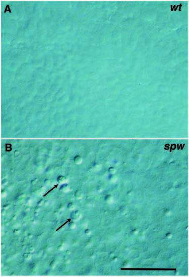
Neuroepithelial tissues at 24 hpf. (A) Wild-type. (B) Morphology of dying cells in space cowboym332. Arrows indicate dying cells. Cells viewed with DIC optics. Scale bar, 25 µm. PHENOTYPE:
|
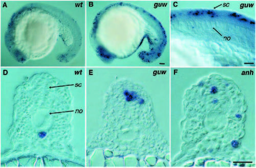
DNA fragmentation in 20-somite stage embryos. (A,D) Wild-type. (B,C,E) gumowym585. DNA fragmentation is localized to the spinal cord and anterior neuroectoderm in mutant embryos. (F) anhalterm767. DNA fragmentation is increased in all major embryonic tissues. (DF) Transverse sections at trunk level. Cells undergoing DNA fragmentation are stained blue. no, notochord; sc, spinal cord. Scale bars, (A,B) 100 µm, (C) 25 µm, (D-F) 25 µm. |
Head phenotypes of miscellaneous early degeneration mutants. Dorsal view onto brain of 20-somite stage embryos. (A) Wild-type. (B) stop and dropm367. (C) sideswipem762. (D) hörnlem274. (E) anhalterm767. Living embryos viewed with DIC optics, anterior to the left. Scale bar, 100 µm. PHENOTYPE:
|
DNA fragmentation in miscellaneous early degeneration mutants at the 20-somite stage. (A,B) Wild-type. (C,D) stop and dropm367. (E,F) sideswipem762. (G,H) hörnlem274. (I,J) anhalterm767. (A,C,E,G,I) Lateral view of live embryos, viewed with DIC optics, anterior to the left. (B,D,F,H,J) Whole-mount staining for DNA fragmentation. Scale bars, 100 µm. |
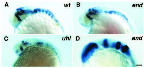
Expression patterns of region-specific markers in spacehead mutant anterior neuroectoderm. (A-C) hlx-1 expression at 28 hpf. (A) Wild-type. (B) endeavorm591. (C) uchu hikoushim172. (D) Expression pattern of pax a and b and krx-20 in endeavorm591 at the 10-somite stage is normal. Lateral view. Viewed with DIC optics, anterior to the left. Scale bar, 100 µm. EXPRESSION / LABELING:
PHENOTYPE:
|
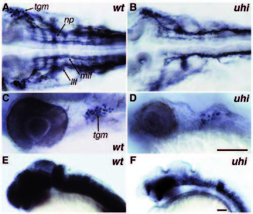
Analysis of neural cell types in mutant uchu hikoushim172 at 30 hpf. (A,C,E) Wild-type. (B,D,F) uchu hikoushim172. (A,B) Dorsal view of znp-1 antibody staining pattern in hindbrain region. Neuropils in each hindbrain segment are affected in mutants. (C,D) Lateral view of Islet-1 antibody staining pattern caudal to the eye. The number of trigeminal ganglion cells is reduced in mutant embryos. (E,F) Lateral view of zrf-1 antibody staining pattern in head region. Mutant embryos have severly reduced amounts of radial glia cells. np, neuropil in hindbrain segment; llf, lateral longitudinal fascicle; mlf, medial longitudinal fascicle; tgm, trigeminal ganglion. Scale bars, (A-D), 100 µm; (E,F) 100 µm. |
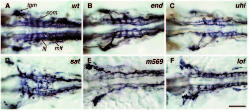
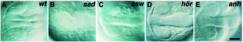
Staining pattern of acetylated tubulin antibody in hindbrain region of spacehead and fala-like classes of mutants at 28 hpf. (A) Wild-type. Normal ladder array of longitudinal fascicles with commissural tracts in each hindbrain segment. (B) endeavorm591. Mild phenotype. (C) uchu hikoushim172. (D) saturnm465. (E) m569. (F) ,em>lost and foundm236. (B-F) Variability of mutant phenotypes. Longitudinal fascicles and commissural tracts are less fasciculated in mutants. Commissural tracts appear to be diminished or to cross the midline in aberrant positions. Dorsal view onto hindbrain. Viewed with DIC optics, anterior to the left. com, commissural tract; llf, lateral longitudinal fascicle; mlf, medial longitudinal fascicle; tgm, trigeminal ganglion. Scale bar, 100 µm. |