
FIGURE
Fig. 5
Fig. 5
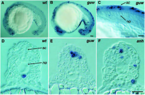
DNA fragmentation in 20-somite stage embryos. (A,D) Wild-type. (B,C,E) gumowym585. DNA fragmentation is localized to the spinal cord and anterior neuroectoderm in mutant embryos. (F) anhalterm767. DNA fragmentation is increased in all major embryonic tissues. (DF) Transverse sections at trunk level. Cells undergoing DNA fragmentation are stained blue. no, notochord; sc, spinal cord. Scale bars, (A,B) 100 µm, (C) 25 µm, (D-F) 25 µm. |
Expression Data
Expression Detail
Antibody Labeling
Phenotype Data
| Fish: | |
|---|---|
| Observed In: | |
| Stage: | 20-25 somites |
Phenotype Detail
Acknowledgments
This image is the copyrighted work of the attributed author or publisher, and
ZFIN has permission only to display this image to its users.
Additional permissions should be obtained from the applicable author or publisher of the image.
Full text @ Development