
- Title
-
The Lysosomal Transcription Factor TFEB Represses Myelination Downstream of the Rag-Ragulator Complex
- Authors
- Meireles, A.M., Shen, K., Zoupi, L., Iyer, H., Bouchard, E.L., Williams, A., Talbot, W.S.
- Source
- Full text @ Dev. Cell
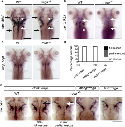
rraga Is Essential for CNS Myelination and Acts Autonomously in Oligodendrocytes (A–E) Analysis of mbp or plp1b mRNA expression at 5 dpf by whole mount in situ hybridization. (A) Compared to their wild-type siblings, rraga−/− mutants show reduced mbp expression in the CNS (white arrows), whereas mbp expression in the PNS is normal (black arrows). (B) Expression of plp1b is also reduced in rraga−/− mutants. See also Figure S1. (C) mtor−/− mutants are developmentally delayed and have a small reduction in mbp mRNA expression levels in the CNS and PNS. See also Figure S2. (D) Quantification of rescue of mbp expression in rraga−/−mutants following expression of wild-type rraga under the control of the claudink, mpeg (expressed in macrophages), or huc promoter (expressed in neurons). (E) Whole mount in situ hybridization analysis of mbp mRNA expression in 5-dpf rraga−/− mutants following expression of wild-type rraga under the control of different tissue-specific promoters. mbp mRNA expression is partially or fully rescued only when wild-type rraga is expressed in oligodendrocytes (claudink promoter). All panels show dorsal views, with anterior to the top. Genotypes of all animals shown were determined by PCR after imaging. Scale bar, 50 μm. |
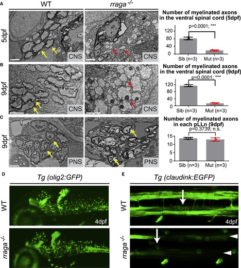
CNS Myelination Is Severely Reduced in rraga−/− Mutants although Oligodendrocytes Are Present (A and B) TEM images of transverse sections of the ventral spinal cord (CNS) at 5 dpf (A) and 9 dpf (B) show fewer myelinated axons in rraga−/− mutants (middle panel) compared to wild-type siblings (left panel). Quantification of the number of myelinated axons in the ventral spinal cord at 5 dpf and 9 dpf is shown on the right; bar graph depicts average values and standard error; individual measurements are also shown. (C) TEM images of transverse sections of the posterior lateral line nerve (pLLn) at 9 dpf show normal myelination in rraga−/− mutants (middle panel) compared to the wild-type sibling (left panel). Quantification of the number of myelinated axons in the pLLn at 9 dpf is shown on the right; bar graph depicts average values and standard error; individual measurements are also shown. In A–C, red arrows indicate unmyelinated axons, and yellow arrows indicate myelinated axons. 3 wild-type and 3 rraga−/− mutants were analyzed. Scale bar, 1 μm. (∗∗∗p < 0.001; Student’s t test, two-tailed). (D) Lateral view of 4 dpf Tg(olig2:EGFP) larvae showing comparable numbers of olig2-positive cells in both the wild-type (top panel) and rraga−/− mutant (lower panel); panel shows anterior to the left and dorsal up. See also Figure S2. (E) Dorsal view of 4-dpf Tg(claudink:EGFP) embryos showing presence of oligodendrocytes (arrows) in both wild-type (top panel) and rraga−/− mutants (lower panel) but reduced expression of GFP along myelinated axonal tracts (arrowheads). Panel shows anterior to the left and dorsal up. Genotypes of all animals shown were determined by PCR after imaging. EXPRESSION / LABELING:
PHENOTYPE:
|
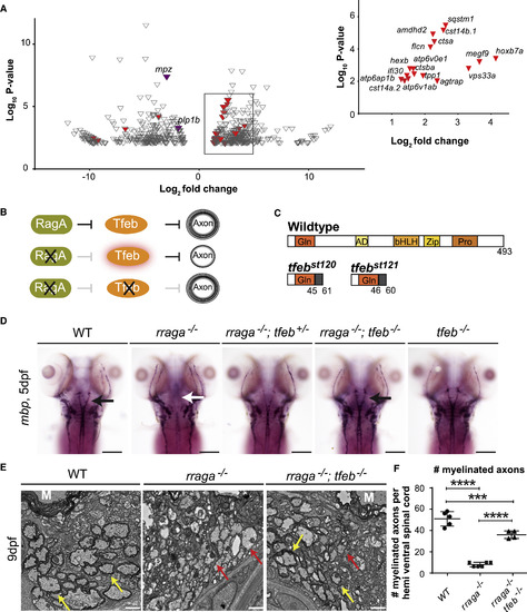
RagA Promotes CNS Myelination by Repressing Tfeb (A) Volcano plot depicting the genes differentially expressed (p < 0.01) in rraga−/− mutants relative to wild-type animals. Among the significantly downregulated genes (< 2.5×) are mpz and plp1b (purple), which at this stage are characteristic of CNS myelin. Expression levels of 17 previously defined targets of TFEB (red) are upregulated (> 2.5) in rraga−/− mutants. Inset graph depicts previously known TFEB targets (red) upregulated in rraga−/− mutants. See also Tables S1, S2, and S3. (B) Diagrammatic representation of the hypothesis that RagA promotes myelination by repressing Tfeb, which inhibits CNS myelination. (C) Diagrammatic representation of Tfeb protein and of predicted truncated proteins encoded by tfebst120 and tfebst121 mutant alleles. (D) Expression of mbp mRNA, as detected by whole mount in situ hybridization, is reduced in CNS of rraga−/− mutants, but is restored in rraga−/−;tfeb−/− double mutants. rraga−/−;tfeb−/− double mutants and tfeb−/− mutants are indistinguishable from wild-type larvae. See also Figure S5. Scale bar, 50 μm. (E) TEM images of transverse sections of the ventral spinal cord (CNS) at 9 dpf show that myelination is restored in rraga−/−;tfeb−/− double mutants. Red arrows indicate unmyelinated axons, and yellow arrows indicate myelinated axons. Scale bar, 1μm. (F) Quantification of the number of myelinated axons per hemi ventral spinal cord at 9 dpf is shown on the right; graph depicts average values and standard deviation; individual measurements are also shown. 3 individuals of each genotype were analyzed. (Statistical analysis: pairwise comparisons using one-way ANOVA; Tukey post hoc test significant interaction ∗∗∗∗p < 0.0001, ∗∗∗p < 0.001). |
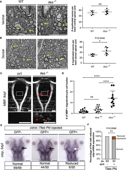
TFEB Represses Myelination in the CNS (A and B) TEM images of transverse sections of the spinal cord at 9 dpf show an increased number of myelinated axons in the dorsal spinal cord of tfeb mutants. Quantification of the number of myelinated axons per ventral (A) or dorsal (B) hemi spinal cord is shown on the right. Graph depicts average values and standard deviation; individual measurements are also shown. Yellow arrows indicate myelinated axons. Scale bar, 5 μm. (Statistical analysis: two-tailed unpaired t test with Welch’s correction). 3 wild-type and 4 tfeb−/− mutant animals were analyzed. (C) Dorsal view of the hindbrain of 8-dpf larvae shows robust expression of MBP protein in myelinating oligodendrocyte processes in wild-type and tfeb−/− mutants and ectopic expression of MBP protein in tfeb−/− mutant cell bodies (red arrows). Scale bar, 50 μm. (D) Quantification of the number of ectopic MBP-positive cells in tfeb−/− mutants (tfeb−/−; n = 11) and siblings (n = 13 for wild-type and tfeb+/− heterozygotes) (Pairwise comparisons using one-way ANOVA; Tukey post hoc test significant interaction; ∗∗∗∗p < 0.0001); bars represent standard deviation values. Individual values are also depicted. Genotypes of all animals shown were determined by PCR after imaging. (E) Whole mount in situ hybridization analysis of mbp mRNA expression in 5-dpf fish following transgenic expression of phosphorylation-null Tfeb (Tfeb NP) under the control of claudink promoter. mbp mRNA expression is reduced in some fish overexpressing the construct. (F) Quantification of the percentage of fish with reduced mbp mRNA expression. (Statistical analysis: Fisher’s exact test, two-tailed, ∗∗p < 0.01). |
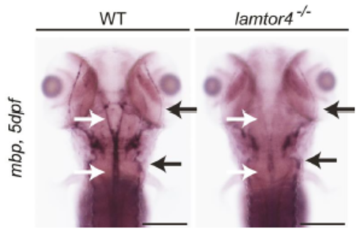
lamtor4 is essential for CNS myelination. (A) Analysis of mbp mRNA expression at 5 dpf by whole mount in situ hybridization. Compared to their wildtype siblings, lamtor4-/- mutants show reduced mbp expression in the CNS (white arrows), whereas mbp expression in the PNS is normal (black arrows). All panels show dorsal views, with anterior to the top. Genotypes were determined by PCR after imaging. Scale bar = 50 μm. |

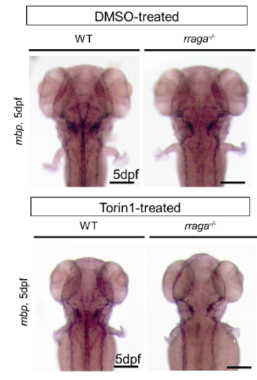
Torin treated fish are developmentally delayed mbp mRNA expression at 5 dpf as detected by whole mount in situ hybridization. Wildtype larvae treated with the mTOR signaling pathway inhibitor TORIN1 have delayed developmental progression and slightly reduced levels of mbp mRNA both in the CNS and PNS. Genotypes were determined by PCR after imaging. Scale bar = 50 μm. |
rraga-/- mutants have normal number of cells expressing olig2. (A) Analysis of olig2 mRNA expression at 3 dpf by whole mount in situ hybridization. Number of olig2 positive cells is similar in rraga-/- mutants and wildtype siblings. (B) Dorsal view of 4 dpf Tg(claudinK:GFP) zebrafish larvae shows that rraga-/- mutants express claudink:GFP, but at reduced levels. Genotypes were determined by PCR after imaging |
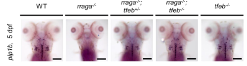
plp expression is restored in rraga-/-;tfeb-/- double mutants Dorsal view of 5 dpf whole mount in situ hybridization to detect plp1b mRNA. Expression of plp1b mRNA is nearly absent in rraga-/- mutants, but is restored in rraga-/-;tfeb-/- double mutants. By this assay rraga-/-;tfeb-/- double mutants and tfeb-/- mutants are indistinguishable from wildtype larvae. Expression of mbp is also partly rescued in rraga-/-;tfeb+/- mutants. Genotypes were determined by PCR after imaging. Scale bar = 50 μm |
Reprinted from Developmental Cell, 47, Meireles, A.M., Shen, K., Zoupi, L., Iyer, H., Bouchard, E.L., Williams, A., Talbot, W.S., The Lysosomal Transcription Factor TFEB Represses Myelination Downstream of the Rag-Ragulator Complex, 319-330.e5, Copyright (2018) with permission from Elsevier. Full text @ Dev. Cell