
- Title
-
Neuronal cell culture from transgenic zebrafish models of neurodegenerative disease
- Authors
- Acosta, J.R., Watchon, M., Yuan, K.C., Fifita, J., Svahn, A.J., Don, E.K., Blair, I.P., Nicholson, G.A., Cole, N.J., Goldsbury, C., Laird, A.S.
- Source
- Full text @ Biol. Open
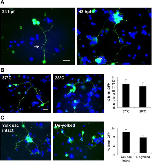
Optimization of the zebrafish primary neural cell culture. (A) Images of cell cultures derived from 24 hpf and 48 hpf-aged embryos. Motor neurons in both cultures exhibited outgrowth of long processes (arrows). (B) No difference in motor neuron survival rate was evident for cells incubated at 37°C or 28°C after 1 day (cultures from 24 hpf larvae). (C) Motor neurons in cultures derived from de-yolked embryos exhibited shorter neurites compared to those derived from whole embryos. Note however that by 2 div, almost 100% of cells from the de-yolked cultures were non-viable (not shown). Scale bar: 10 µm. |
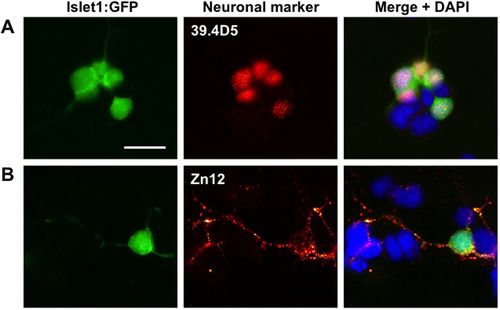
Images of cultured 24 hpf Islet1:GFP zebrafish embryos stained with zebrafish-specific neuronal markers to confirm that the cell cultures contain various types of neurons. (A) An Islet1:GFP motor neuron within the cultures is positively stained (red) for the neuronal marker 39.4D5 (islet1 and islet2 homeobox). (B) Another Islet1:GFP motor neuron, and nearby islet1:GFP negative cells, are stained positively (red) for the neuronal cell surface marker Zn12, indicating the inclusion of other types of neurons in addition to motor neurons. Scale bar: 10 µm. |
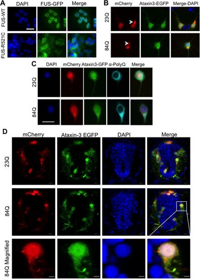
Cultured cells derived from transgenic zebrafish larvae expressing neurodegenerative disease associated proteins FUS or ataxin-3. (A) In cells cultured from mutant human FUS-GFP (FUS-R521C) zebrafish the FUS-GFP protein was mislocalized to the cytosol, whereas it remained predominantly nuclear in cells cultured from wild-type FUS-GFP zebrafish. (B) Cells cultured from double transgenic zebrafish expressing mCherry (red) and EGFP-ataxin-3-23Q/84Q (green) showed no obvious difference in fluorescent protein distribution in cells expressing non-pathogenic EGFP-ataxin-3-23Q and pathogenic EGFP-ataxin-3-84Q. Aggregates of mCherry-positive protein (arrows) were present in some neurons (Jakobs et al., 2000). (C) Immunolabeling cell cultures with anti-polyQ (pale blue) demonstrated cytosolic distribution of the ataxin-3 protein in cells expressing either EGFP-ataxin-3-23Q or pathogenic EGFP-ataxin-3-84Q. Scale bars: 10 µm. (D) Cross-sections of the spinal cord of 3 dpf transgenic SCA3 zebrafish revealed a similar expression pattern of EGFP-ataxin-3 and mCherry to that seen in the cell cultures. Scale bars: 5 µm. |