
FIGURE
Fig. 1
- ID
- ZDB-FIG-181127-46
- Publication
- Acosta et al., 2018 - Neuronal cell culture from transgenic zebrafish models of neurodegenerative disease
- Other Figures
- All Figure Page
- Back to All Figure Page
Fig. 1
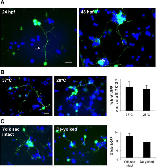
Optimization of the zebrafish primary neural cell culture. (A) Images of cell cultures derived from 24 hpf and 48 hpf-aged embryos. Motor neurons in both cultures exhibited outgrowth of long processes (arrows). (B) No difference in motor neuron survival rate was evident for cells incubated at 37°C or 28°C after 1 day (cultures from 24 hpf larvae). (C) Motor neurons in cultures derived from de-yolked embryos exhibited shorter neurites compared to those derived from whole embryos. Note however that by 2 div, almost 100% of cells from the de-yolked cultures were non-viable (not shown). Scale bar: 10 µm. |
Expression Data
Expression Detail
Antibody Labeling
Phenotype Data
Phenotype Detail
Acknowledgments
This image is the copyrighted work of the attributed author or publisher, and
ZFIN has permission only to display this image to its users.
Additional permissions should be obtained from the applicable author or publisher of the image.
Full text @ Biol. Open