
- Title
-
Pineal photoreceptor cells are required for maintaining the circadian rhythms of behavioral visual sensitivity in zebrafish
- Authors
- Li, X., Montgomery, J., Cheng, W., Noh, J.H., Hyde, D.R., and Li, L.
- Source
- Full text @ PLoS One
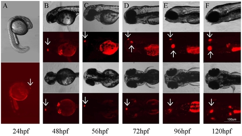
Bright-field and fluorescent images of developing embryos at different developmental stages that show Tg(Gnat2:gal4-VP16/UAS:nfsB-mCherry) transgene expression in the pineal gland and retina. (A) At 24 hpf, mCherry was detected in the pineal gland (arrow). (B) Strong mCherry expression was seen in the pineal gland (arrows). (C–F) Between 56 and 120 hpf, mCherry was detected in both the pineal gland (downward arrows) and retina (upward arrows). The intensity of transgene expression increased during embryonic development. |
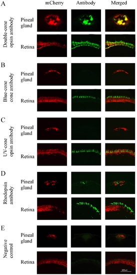
Cryostat sections of pineal gland and retina of transgenic zebrafish (2-month old) that show mCherry (left panels), opsin antibody immunoreactivity (middle panels), and co-localization of mCherry and opsin antibodies (right panels). (A) mCherry and double-cone opsin immunoreactivity in the pineal gland and retina. mCherry and double-cone opsin antibodies were co-localized in the pineal and retinal photoreceptor cells. (B, C) mCherry, blue- and UV-cone opsin immunoreactivity in the pineal gland and retina. Blue-cone and UV-cone antibody immunoreactivity were not seen in the pineal gland, but were detected in the retina where they co-localized with mCherry. (D) mCherry and rhodopsin immunoreactivity in the pineal gland and retina. Rhodopsin antibody immunoreactivity was detected in both pineal and retinal rod photoreceptor cells, but did not co-localize with mCherry. (E) Fluorescent images of pineal and retinal sections that were processed without primary antibodies (negative controls). |
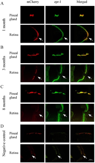
Cryostat sections of pineal gland and retina of transgenic zebrafish (1, 3 and 8 months) that show mCherry (left panels), double-cone opsin antibody (zpr-1) immunoreactivity (middle panels), and co-localization of mCherry and double-cone opsin antibody (right panels). (A) At 1 month of age, strong transgene expression was seen in the pineal gland and retina. mCherry and zpr-1 immunoreactivity were co-localized (B) At 3 months, the expression of mCherry decreased in the retina, but increased in the pineal gland. Co-localized of mCherry and zpr-1 immunoreactivity was detected in the pineal gland and central retina. (C) By 8 months, mCherry was detected in the pineal gland, but not in the retina. (D) Fluorescent images of pineal and retinal sections (from 8-months-old animals) that were processed without primary antibodies (negative controls). Arrows point the optic nerve. |
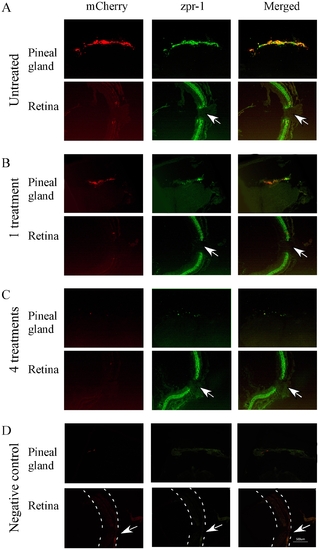
Cryostat sections of pineal gland and retina of transgenic zebrafish (8-month old) that show mCherry (left panels), double-cone opsin antibody immunoreactivity (zpr-1, middle panels), and co-localization of mCherry and opsin antibody (right panels) before and after the treatment with metronidazole. (A) Pineal and retinal sections from untreated transgenic fish. Strong mCherry expression was detected in the pineal gland, but not in the retina. zpr-1 immunoreactivity was seen in both pineal and retinal photoreceptor cells. (B) After 1 metronidazole treatment, mCherry expression and zpr-1 immunoreactivity in the pineal gland were decreased. In the retina, strong zpr-1 immunoreactivity was detected. (C) After 4 metronidazole treatments, the expression of mCherry and zpr-1 immunoreactivity in the pineal gland was completely diminished. Strong zpr-1 immunoreactivity was detected in the retina. (D) Fluorescent images of pineal and retinal sections that were processed without primary antibodies (negative controls). Dashed lines outline the retina. Arrows point to the optic nerve. |
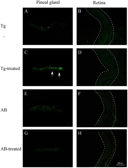
TUNEL labeling of pineal and retinal sections of metronidazole-treated and untreated transgenic (Tg) and wild-type (AB) zebrafish (8 months old). (A, B) Pineal and retinal sections of untreated transgenic fish. No obvious cell death was detected. (C, D) Pineal and retinal sections of transgenic zebrafish after 4 metronidazole treatments. In the pineal gland, TUNEL-positive cells were detected (arrows). No cell death was seen in the retina. (E–H) Pineal and retinal sections from untreated and metronidazole-treated wild-type zebrafish. No cell death was seen in the pineal gland or retina. Dashed lines outline the retina. |
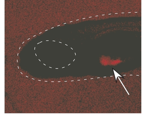
A wild-type embryo at 3 dpa. The arrow points the yolk that shows auto-fluorescence. Outlines indicate the head and eye. |

DAPI staining of the pineal gland of untreated (A) and metronidazole-treated (B) transgenic zebrafish at 8 months old. Note the decrease in DAPI staining after metronidazole treatment. |